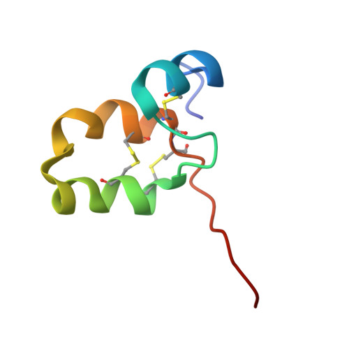
37EI | pdb_000037ei

Solution structure of the Cys-rich domain of the 15 kDa selenoprotein

  • Classification: CHAPERONE
  • Organism(s): Homo sapiens
  • Expression System: Escherichia coli
  • Mutation(s): No 

  • Deposited: 2026-07-16 Released: 2026-10-28 
  • Deposition Author(s): Williams, R., Gierasch, L.
  • Funding Organization(s): National Institutes of Health/National Institute of General Medical Sciences (NIH/NIGMS)

Experimental Data Snapshot

  • Method: SOLUTION NMR
  • Conformers Calculated: 50 
  • Conformers Submitted: 20 
  • Selection Criteria: structures with the lowest energy 

wwPDB Validation 3D Report Full Report

Validation slider image for 37EI

This is version 1.0 of the entry. See complete history. 

Literature

The cysteine-rich domain of SEP15, a selenoprotein co-chaperone of the ER chaperone, UDP-glucose:glycoprotein glucosyltransferase, adopts a novel fold.

Williams, R.V., Guay, K.P., Lesk, O.H., Hebert, D.N., Gierasch, L.M.

(2026) J Biol Chem : 113629-113629

  • DOI: https://doi.org/10.1016/j.jbc.2026.113629
  • Primary Citation Related Structures: 
    37EI, 9A9H

  • PubMed Abstract: 

    Proteins targeted to the secretory pathway are involved in a myriad of biological processes but can only do so when properly folded. Within the endoplasmic reticulum, glycoprotein folding is regulated by the enzyme UDP-glucose:glycoprotein glucosyltransferase (UGGT) and its oxidoreductase partner, the 15-kDa selenoprotein (SEP15 aka SELENOF). The interaction between these two chaperones is poorly understood, limiting understanding of their function. SEP15 is comprised of two domains, a C-terminal thioredoxin-like domain, the structure of which has been reported (PDB 2A4H), and an approximately 50-residue long N-terminal cysteine-rich domain (CRD), of unknown structure. Here, we report the NMR solution structure of SEP15 CRD, which mediates the interaction with UGGT. These data reveal that this domain adopts a dimer in solution and forms a previously undescribed helical fold, stabilized by three disulfide bonds between residues C10-C42, C21-C43, and C24-C39. Furthermore, our results validate a model of the UGGT/SEP15 complex and lay the foundation for future studies of its interaction with glycoprotein substrates.


  • Organizational Affiliation: 
    • Department of Biochemistry & Molecular Biology, University of Massachusetts Amherst, Amherst, MA 01003.

Macromolecule Content 

  • Total Structure Weight: 11.49 kDa 
  • Atom Count: 796 
  • Modeled Residue Count: 104 
  • Deposited Residue Count: 104 
  • Unique protein chains: 1

Macromolecules

Find similar proteins by:|  3D Structure
Entity ID: 1
MoleculeChains  Sequence LengthOrganismDetailsImage
Selenoprotein F
A, B
52Homo sapiensMutation(s): 0 
Gene Names: SELENOF, SEP15
UniProt & NIH Common Fund Data Resources
Find proteins for O60613 (Homo sapiens)
Explore O60613 
Go to UniProtKB:  O60613
PHAROS:  O60613
GTEx:  ENSG00000183291 
Entity Groups
Sequence Clusters30% Identity50% Identity70% Identity90% Identity95% Identity100% Identity
UniProt GroupO60613
Sequence Annotations
Expand
Reference Sequence

Experimental Data & Validation

Experimental Data

  • Method: SOLUTION NMR
  • Conformers Calculated: 50 
  • Conformers Submitted: 20 
  • Selection Criteria: structures with the lowest energy 

Structure Validation

View Full Validation Report


Entry History 

& Funding Information

Deposition Data


Funding OrganizationLocationGrant Number
National Institutes of Health/National Institute of General Medical Sciences (NIH/NIGMS)United StatesGM118161

Revision History  (Full details and data files)

  • Version 1.0: 2026-10-28
    Type: Initial release