36JB | pdb_000036jb

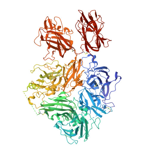
Coagulation factor V DTQQ, membrane-bound, on Ptd-choline : Ptd-serine 75:25 vesicles


Experimental Data Snapshot

  • Method: ELECTRON MICROSCOPY
  • Resolution: 2.90 Å
  • Aggregation State: PARTICLE 
  • Reconstruction Method: SINGLE PARTICLE 

wwPDB Validation 3D Report Full Report

Validation slider image for 36JB

This is version 1.0 of the entry. See complete history

Literature

Structural basis for membrane binding by coagulation factors V and VIII and their specificity for phosphatidylserine-containing membranes.

Kolyadko, V.N.Pumroy, R.A.Moiseenkova-Bell, V.Y.Krishnaswamy, S.

(2026) Proc Natl Acad Sci U S A 123: e2622255123-e2622255123

  • DOI: https://doi.org/10.1073/pnas.2622255123
  • Primary Citation Related Structures: 
    36JA, 36JB, 36JK, 36JL

  • PubMed Abstract: 

    Phosphatidylserine on the surface of activated cells serves as a signal for coagulation factor binding and the membrane-dependent assembly of enzyme complexes that drive the accelerated host response to vascular damage. Here, we use single-particle cryo-EM of liposome-bound proteins to establish common mechanisms utilized by coagulation factors V and VIII for membrane binding and phospholipid specificity. The interface between these peripheral proteins and the membrane shows contacts between the discoidin C1 and C2 domains and the interfacial phospholipid headgroup region of the membrane bilayer. There is no evidence for the insertion of membrane-contacting protein loops into the membrane core. This refutes previous models of high affinity protein binding by insertion into the hydrophobic core of the membrane. We also resolve density for the headgroup of phosphatidylserine bound to conserved pockets within the C1 and C2 domains. These single binding pockets in each C domain highlight the network of putative hydrogen bonds that contribute to binding specificity for phosphatidylserine. Our results now provide a high-resolution structural view of the protein-membrane interface for these coagulation proteins and highlight the utility of using liposomes as near-physiologic membrane mimetics in structural studies. The principles established in this work may also apply to other biological systems wherein function is regulated by reversible membrane binding.


  • Organizational Affiliation
    • Division of Hematology, Department of Pediatrics, Children's Hospital of Philadelphia, Philadelphia, PA 19104.

Macromolecule Content 

  • Total Structure Weight: 176.25 kDa 
  • Atom Count: 10,077 
  • Modeled Residue Count: 1,225 
  • Deposited Residue Count: 1,515 
  • Unique protein chains: 1

Macromolecules

Find similar proteins by:|  3D Structure
Entity ID: 1
MoleculeChains  Sequence LengthOrganismDetailsImage
Coagulation factor V1,515Homo sapiensMutation(s): 2 
Gene Names: F5
UniProt & NIH Common Fund Data Resources
Find proteins for P12259 (Homo sapiens)
Explore P12259 
Go to UniProtKB:  P12259
PHAROS:  P12259
GTEx:  ENSG00000198734 
Entity Groups
Sequence Clusters30% Identity50% Identity70% Identity90% Identity95% Identity100% Identity
UniProt GroupP12259
Glycosylation
Glycosylation Sites: 4Go to GlyGen: P12259-1
Sequence Annotations
Expand
Reference Sequence

Oligosaccharides

Help  
Entity ID: 2
MoleculeChains Length2D Diagram GlycosylationD Interactions
2-acetamido-2-deoxy-beta-D-glucopyranose-(1-4)-2-acetamido-2-deoxy-beta-D-glucopyranose
B, C
2N-Glycosylation
Entity ID: 3
MoleculeChains Length2D Diagram GlycosylationD Interactions
alpha-D-mannopyranose-(1-3)-[alpha-D-mannopyranose-(1-6)]beta-D-mannopyranose-(1-4)-2-acetamido-2-deoxy-beta-D-glucopyranose-(1-4)-2-acetamido-2-deoxy-beta-D-glucopyranose
D
5N-Glycosylation

Small Molecules

Ligands 4 Unique
IDChains Name / Formula / InChI Key2D Diagram3D Interactions
NAG

Query on NAG


Download:Ideal Coordinates CCD File
E [auth A]2-acetamido-2-deoxy-beta-D-glucopyranose
C8 H15 N O6
OVRNDRQMDRJTHS-FMDGEEDCSA-N
SEP
(Subject of Investigation/LOI)

Query on SEP


Download:Ideal Coordinates CCD File
I [auth A],
J [auth A]
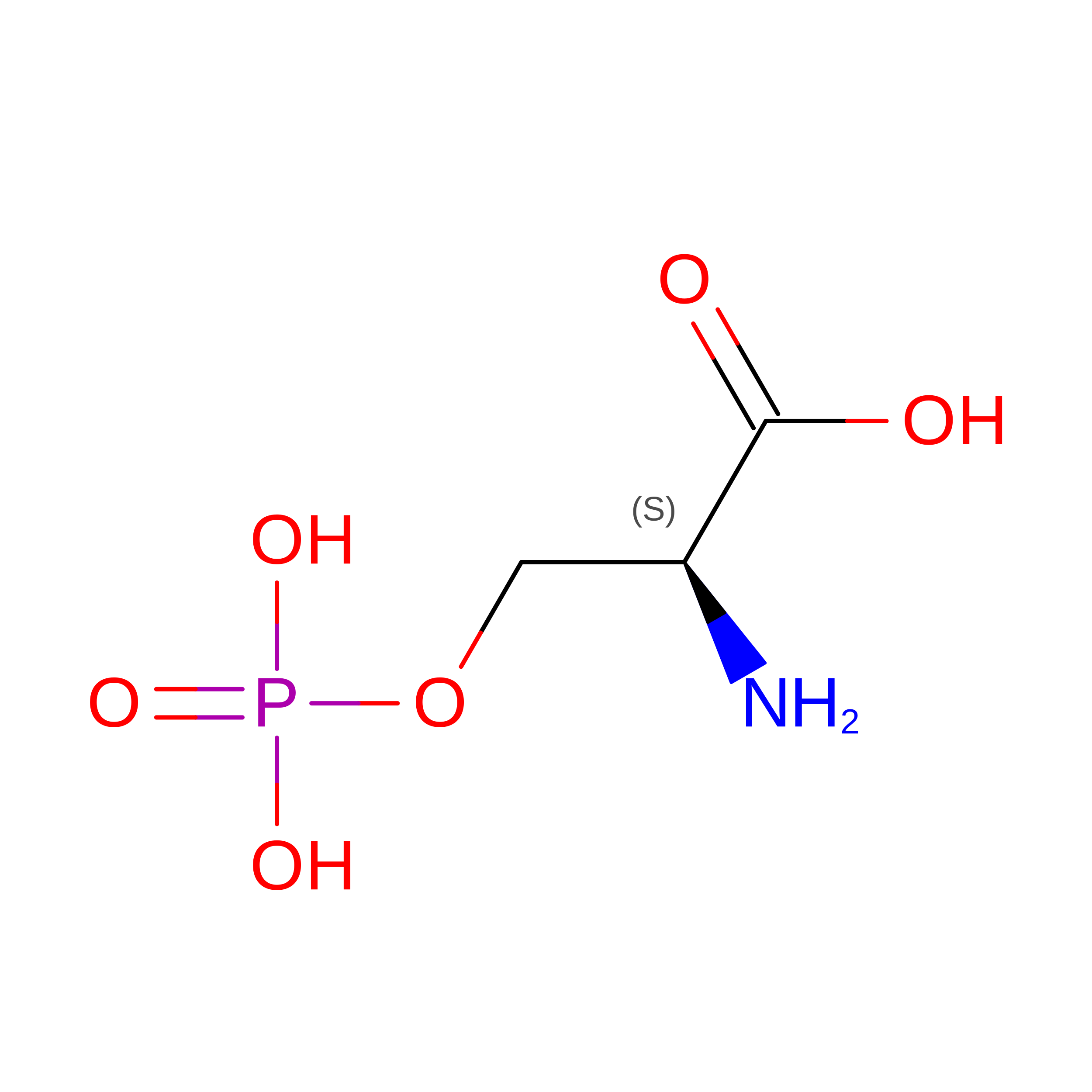
PHOSPHOSERINE
C3 H8 N O6 P
BZQFBWGGLXLEPQ-REOHCLBHSA-N
CU1
(Subject of Investigation/LOI)

Query on CU1


Download:Ideal Coordinates CCD File
H [auth A]COPPER (I) ION
Cu
VMQMZMRVKUZKQL-UHFFFAOYSA-N
CA
(Subject of Investigation/LOI)

Query on CA


Download:Ideal Coordinates CCD File
F [auth A],
G [auth A]
CALCIUM ION
Ca
BHPQYMZQTOCNFJ-UHFFFAOYSA-N

Experimental Data & Validation

Experimental Data

  • Method: ELECTRON MICROSCOPY
  • Resolution: 2.90 Å
  • Aggregation State: PARTICLE 
  • Reconstruction Method: SINGLE PARTICLE 
EM Software:
TaskSoftware PackageVersion
MODEL REFINEMENTPHENIX1.20.1_4487
RECONSTRUCTIONcryoSPARC4.6.1

Structure Validation

View Full Validation Report


Entry History 

& Funding Information

Deposition Data


Funding OrganizationLocationGrant Number
National Institutes of Health/National Heart, Lung, and Blood Institute (NIH/NHLBI)United StatesP01HL139420

Revision History  (Full details and data files)

  • Version 1.0: 2026-09-16
    Type: Initial release