 Download Mendeley
Defining Glycosyl Hydrolase Family 5_7 mannan interaction through the identification and structural characterization of two Actinobacteria mannanases.
Bloch, Y.To be published.
Experimental Data Snapshot
Starting Model: experimental
View more details
wwPDB Validation 3D Report Full Report

Entity ID: 1 | |||||
|---|---|---|---|---|---|
| Molecule | Chains | Sequence Length | Organism | Details | Image |
| GH5_7 Mannanase | 398 | Streptomyces sp. TLI_171 | Mutation(s): 0 EC: 3.2.1.78 | 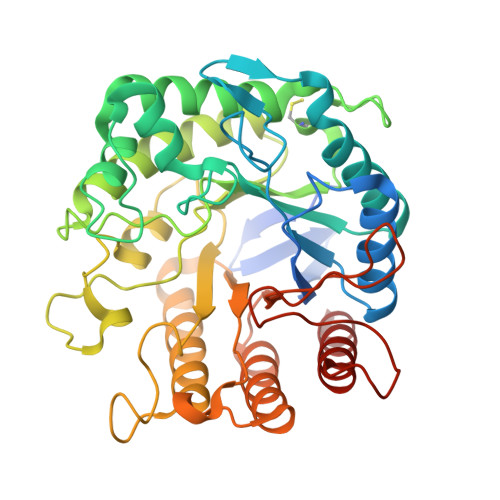 | |
Entity Groups | |||||
| Sequence Clusters | 30% Identity50% Identity70% Identity90% Identity95% Identity100% Identity | ||||
Sequence AnnotationsExpand | |||||
Reference Sequence | |||||
Entity ID: 2 | |||||
|---|---|---|---|---|---|
| Molecule | Chains | Sequence Length | Organism | Details | Image |
| GH5_7 Mannanase | 398 | Streptomyces sp. TLI_171 | Mutation(s): 0 EC: 3.2.1.78 | 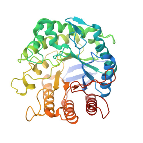 | |
Entity Groups | |||||
| Sequence Clusters | 30% Identity50% Identity70% Identity90% Identity95% Identity100% Identity | ||||
Sequence AnnotationsExpand | |||||
Reference Sequence | |||||
Entity ID: 3 | |||||
|---|---|---|---|---|---|
| Molecule | Chains | Length | 2D Diagram | Glycosylation | D Interactions |
| beta-D-mannopyranose-(1-4)-beta-D-mannopyranose-(1-4)-beta-D-mannopyranose-(1-4)-beta-D-mannopyranose-(1-4)-beta-D-mannopyranose-(1-4)-beta-D-mannopyranose | E, F | 6 |  | N/A | |
Entity ID: 4 | |||||
|---|---|---|---|---|---|
| Molecule | Chains | Length | 2D Diagram | Glycosylation | D Interactions |
| beta-D-mannopyranose-(1-4)-beta-D-mannopyranose-(1-4)-beta-D-mannopyranose-(1-4)-beta-D-mannopyranose-(1-4)-beta-D-mannopyranose | G | 5 |  | N/A | |
Entity ID: 5 | |||||
|---|---|---|---|---|---|
| Molecule | Chains | Length | 2D Diagram | Glycosylation | D Interactions |
| beta-D-mannopyranose-(1-4)-beta-D-mannopyranose-(1-4)-beta-D-mannopyranose-(1-4)-beta-D-mannopyranose-(1-4)-beta-D-mannopyranose-(1-4)-beta-D-mannopyranose-(1-4)-beta-D-mannopyranose | H | 7 |  | N/A | |
| Modified Residues 1 Unique | |||||
|---|---|---|---|---|---|
| ID | Chains | Modified Residue | Modification Type | Formula | 2D Diagram |
| CSS Query on CSS | CYS | Sulfhydration | C3 H7 N O2 S2 |  | |
| Length ( Å ) | Angle ( ˚ ) |
|---|---|
| a = 95.9 | α = 90 |
| b = 106.4 | β = 90 |
| c = 158.02 | γ = 90 |
| Software Name | Purpose |
|---|---|
| PHENIX | refinement |
| XDS | data reduction |
| STARANISO | data scaling |
| PHASER | phasing |
| Funding Organization | Location | Grant Number |
|---|---|---|
| H2020 Marie Curie Actions of the European Commission | European Union | 945405 |
