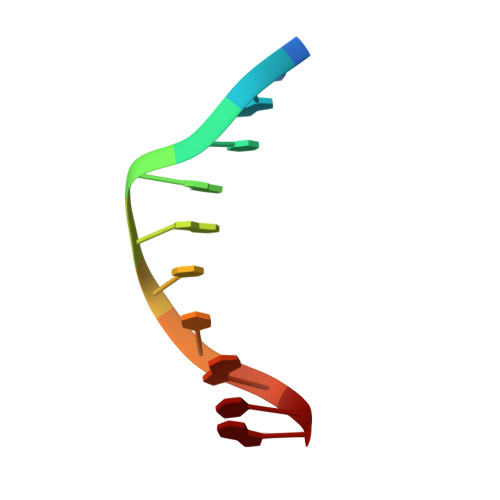
Defining GC-specificity in the minor groove: side-by-side binding of the di-imidazole lexitropsin to C-A-T-G-G-C-C-A-T-G.
Kopka, M.L., Goodsell, D.S., Han, G.W., Chiu, T.K., Lown, J.W., Dickerson, R.E.(1997) Structure 5: 1033-1046
- PubMed: 9309219 Search on PubMed
- DOI: https://doi.org/10.1016/s0969-2126(97)00255-4
- Primary Citation Related Structures:
334D - PubMed Abstract:
Polyamide drugs, such as netropsin, distamycin and their lexitropsin derivatives, can be inserted into a narrow B-DNA minor groove to form 1:1 complexes that can distinguish AT base pairs from GC, but cannot detect end-for-end base-pair reversals such as TA for AT. In contrast, 2:1 side-by-side polyamide drug complexes potentially are capable of such discrimination. Imidazole (Im) and pyrrole (Py) rings side-by-side read a GC base pair with the Im ring recognizing the guanine side. But the reason for this specific G-Im association is unclear because the guanine NH2 group sits in the center of the groove. A 2:1 drug:DNA complex that presents Im at both ends of a GC base pair should help unscramble the issue of imidazole reading specificity. We have determined the crystal structure of a 2:1 complex of a di-imidazole lexitropsin (DIM), an analogue of distamycin, and a DNA decamer with the sequence C-A-T-G-G-C-C-A-T-G. The two DIM molecules sit antiparallel to one another in a broad minor groove, with their cationic tails widely separated. Im rings of one drug molecule stack against amide groups of the other. DIM1 rests against nucleotides C7A8T9G10 of strand 1 of the helix, whereas DIM2 rests against G14G15C16C17 on strand 2. All DIM amide nitrogens donate hydrogen bonds to N and O atoms on the floor of the DNA groove and, in addition, the two Im rings on DIM2 accept hydrogen bonds from guanine N2 amines, thereby providing specific reading. The guanine N2 amine can bond to Im on its own side of the groove, but not on the cytosine side, because of limits on close approach of the two Im rings and the geometry of sp2 hybridization about the amide nitrogen. Im and Py rings distinguish AT from GC base pairs because of steric factors involving the bulk of the guanine amine, and the ability of Im to form a hydrogen bond with the amine. Side-by-side Im and Py rings differentiate GC from CG base pairs because of tight steric contacts and sp2 hybridization at the amine nitrogen atom, with the favored conformations being G/Im,Py/C and C/Py,Im/G. Discrimination between AT and TA base pairs may be possible using bulkier rings, such as thiazole to select the A end of the base pair.
- Molecular Biology Institute, University of California at Los Angeles 90095, USA. kopka@ewald.mbi.ucla.edu
Organizational Affiliation: