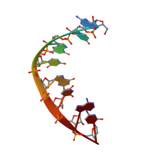
Crystal structure of r(GUGUGUA)dC with tandem G x U/U x G wobble pairs with strand slippage.
Biswas, R., Sundaralingam, M.(1997) J Mol Biology 270: 511-519
- PubMed: 9237915 Search on PubMed
- DOI: https://doi.org/10.1006/jmbi.1997.1118
- Primary Citation Related Structures:
332D - PubMed Abstract:
To better understand the frequent occurrence of adjacent wobble pairs in ribosomal RNAs we have determined the crystal structure of the RNA duplex, r(GUGUGUA)dC with the 3'-terminal deoxy C residue. Two different crystal forms of the duplex were obtained and both belong to the rhombohedral space group, R3. Crystal form I has hexagonal unit cell dimensions, a = b = 40.82 A and c = 66.09 A and diffracts to 1.58 A resolution, while crystal form II has a = b = 47.11 A and c = 59.86 A, diffracting only to 2.50 A resolution. Both structures were solved by the molecular replacement method using different starting models. In spite of the large differences in the cell dimensions the overall structures in both crystals are similar. Instead of the expected blunt-end duplex with four consecutive G x U pairs, the slippage of the strands resulted in two different tandem G x U/U x G wobble pairs involving two of the central and two of the 5' overhang bases, still yielding a total of four wobble pairs. These tandem wobble pairs are flanked by two Watson-Crick pairs. The A-type duplexes stack in the familiar head-to-tail fashion forming a pseudocontinuous helix. The wobble pairs of the present motif II (G x U/U x G) structure stack with a low twist angle of 25.3 degrees in contrast to that of motif I (U x G/G x U), 38.1 degrees. The four wobble pairs are characteristically heavily hydrated in both the grooves accounting for their stability.
- Biological Macromolecular Structure Center, Department of Chemistry, The Ohio State University, Columbus 43210, USA.
Organizational Affiliation: