31AK | pdb_000031ak

Structure of native human leukocyte myeloperoxidase


Experimental Data Snapshot

  • Method: ELECTRON MICROSCOPY
  • Resolution: 2.18 Å
  • Aggregation State: PARTICLE 
  • Reconstruction Method: SINGLE PARTICLE 

wwPDB Validation 3D Report Full Report

Validation slider image for 31AK

This is version 1.0 of the entry. See complete history. 

Literature

Interface N-glycans drive myeloperoxidase dimerization in vitro.

Leitgeb, U., Guo, Y., Emde, T., Ruocco, V., Simak, T., Zdenkovic, E., Furtmuller, P.G., Borek, D., Nauseef, W.M., Oostenbrink, C., Pfanzagl, V.

(2026) Int J Biol Macromol : 154499-154499

  • DOI: https://doi.org/10.1016/j.ijbiomac.2026.154499
  • Primary Citation Related Structures: 
    29TO, 29WJ, 31AK

  • PubMed Abstract: 

    Myeloperoxidase (MPO) is a highly glycosylated heme oxidoreductase that, together with lactoperoxidase (LPO) and eosinophil peroxidase (EPO), contributes to host defence through the generation of (pseudo)hypohalous acids. Among the human heme peroxidases, mature MPO forms a unique covalently linked homodimer. Dimerization enhances stability and is essential for some biological functions, including chromatin disruption during neutrophil extracellular trap formation. Thus, we hypothesized that MPO dimerization must reflect a tightly regulated step in MPO biosynthesis and is driven by specific structural elements. Cross-species sequence alignment indicated that N-glycosylation motifs were evolutionarily more conserved in MPO than in monomeric EPO and LPO. We investigated the role of N-glycans in MPO dimerization using in vitro monomerized glycosylated (mMPO) and deglycosylated (mMPOdg) monomeric variants of native human MPO. Small-angle X-ray scattering, isothermal titration calorimetry and crystal structures showed that glycosylated MPO monomers had a weak but biologically relevant affinity in the millimolar range, which was lost upon deglycosylation. Furthermore, we present the first cryogenic transmission electron microscopy structure of human MPO with at least one core N-acetylglucosamine residue resolved at each N-glycosylation site. Structural analysis suggests that an extensive hydrogen bonding network of interface N-glycans at position 483 and residues of the other monomer drives MPO homodimerization. Dimerization enhances stability without affecting enzymatic activity. Together, we show that N-glycans are essential to MPO dimerization, provide important insights into the impact of interface N-glycans on biophysical and biochemical properties of MPO and discuss the implications for the biological roles of MPO in health and disease.


  • Organizational Affiliation: 
    • Institute of Biochemistry, Department of Natural Sciences and Sustainable Resources, BOKU University, Vienna, Muthgasse 18, 1190, Vienna, Austria.

Macromolecule Content 

  • Total Structure Weight: 140.33 kDa 
  • Atom Count: 9,619 
  • Modeled Residue Count: 1,142 
  • Deposited Residue Count: 1,162 
  • Unique protein chains: 2

Macromolecules

Find similar proteins by:|  3D Structure
Entity ID: 1
MoleculeChains  Sequence LengthOrganismDetailsImage
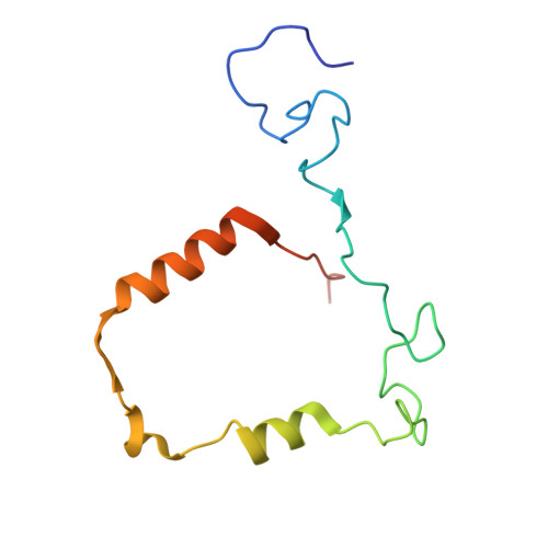
Myeloperoxidase light chainA,
C [auth E]
114Homo sapiensMutation(s): 0 
EC: 1.11.2.2
UniProt & NIH Common Fund Data Resources
Find proteins for P05164 (Homo sapiens)
Explore P05164 
Go to UniProtKB:  P05164
PHAROS:  P05164
GTEx:  ENSG00000005381 
Entity Groups
Sequence Clusters30% Identity50% Identity70% Identity90% Identity95% Identity100% Identity
UniProt GroupP05164
Sequence Annotations
Expand
Reference Sequence
Find similar proteins by:|  3D Structure
Entity ID: 2
MoleculeChains  Sequence LengthOrganismDetailsImage
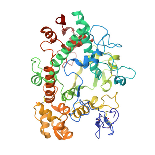
MyeloperoxidaseB,
D [auth F]
467Homo sapiensMutation(s): 0 
EC: 1.11.2.2
UniProt & NIH Common Fund Data Resources
Find proteins for P05164 (Homo sapiens)
Explore P05164 
Go to UniProtKB:  P05164
PHAROS:  P05164
GTEx:  ENSG00000005381 
Entity Groups
Sequence Clusters30% Identity50% Identity70% Identity90% Identity95% Identity100% Identity
UniProt GroupP05164
Glycosylation
Glycosylation Sites: 5Go to GlyGen: P05164-1
Sequence Annotations
Expand
Reference Sequence

Oligosaccharides

Help  
Entity ID: 3
MoleculeChains Length2D Diagram GlycosylationD Interactions
alpha-D-mannopyranose-(1-2)-alpha-D-mannopyranose-(1-3)-[alpha-D-mannopyranose-(1-6)]beta-D-mannopyranose-(1-4)-2-acetamido-2-deoxy-beta-D-glucopyranose-(1-4)-2-acetamido-2-deoxy-beta-D-glucopyranoseE [auth C],
H
6N-Glycosylation
Entity ID: 4
MoleculeChains Length2D Diagram GlycosylationD Interactions
beta-D-mannopyranose-(1-4)-2-acetamido-2-deoxy-beta-D-glucopyranose-(1-4)-2-acetamido-2-deoxy-beta-D-glucopyranoseF [auth D],
I
3N-Glycosylation
Entity ID: 5
MoleculeChains Length2D Diagram GlycosylationD Interactions
alpha-D-mannopyranose-(1-3)-[alpha-D-mannopyranose-(1-6)]beta-D-mannopyranose-(1-4)-2-acetamido-2-deoxy-beta-D-glucopyranose-(1-4)-[alpha-L-fucopyranose-(1-6)]2-acetamido-2-deoxy-beta-D-glucopyranose
G
6N-Glycosylation
Entity ID: 6
MoleculeChains Length2D Diagram GlycosylationD Interactions
alpha-D-mannopyranose-(1-6)-alpha-D-mannopyranose-(1-6)-[alpha-D-mannopyranose-(1-3)]beta-D-mannopyranose-(1-4)-2-acetamido-2-deoxy-beta-D-glucopyranose-(1-4)-[alpha-L-fucopyranose-(1-6)]2-acetamido-2-deoxy-beta-D-glucopyranose
J
7N-Glycosylation

Small Molecules

Ligands 4 Unique
IDChains Name / Formula / InChI Key2D Diagram3D Interactions
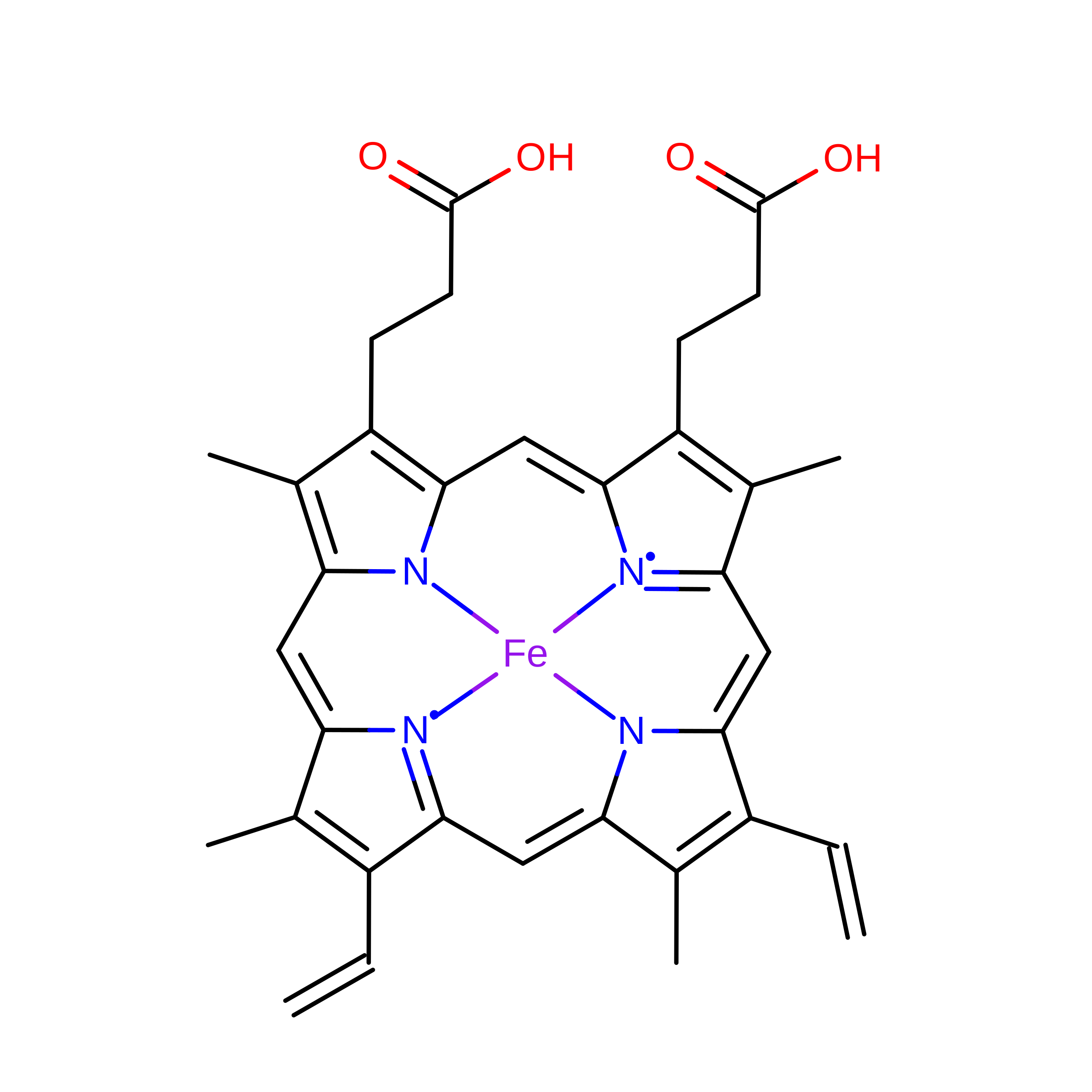
HEM
(Subject of Investigation/LOI)

Query on HEM


Download:Ideal Coordinates CCD File
N [auth B],
R [auth E]
PROTOPORPHYRIN IX CONTAINING FE
C34 H32 Fe N4 O4
KABFMIBPWCXCRK-RGGAHWMASA-L
NAG

Query on NAG


Download:Ideal Coordinates CCD File
O [auth B],
P [auth B],
U [auth F],
V [auth F]
2-acetamido-2-deoxy-beta-D-glucopyranose
C8 H15 N O6
OVRNDRQMDRJTHS-FMDGEEDCSA-N
CA

Query on CA


Download:Ideal Coordinates CCD File
L [auth B],
S [auth F]
CALCIUM ION
Ca
BHPQYMZQTOCNFJ-UHFFFAOYSA-N
CL

Query on CL


Download:Ideal Coordinates CCD File
K [auth A],
M [auth B],
Q [auth E],
T [auth F]
CHLORIDE ION
Cl
VEXZGXHMUGYJMC-UHFFFAOYSA-M

Experimental Data & Validation

Experimental Data

  • Method: ELECTRON MICROSCOPY
  • Resolution: 2.18 Å
  • Aggregation State: PARTICLE 
  • Reconstruction Method: SINGLE PARTICLE 
EM Software:
TaskSoftware PackageVersion
RECONSTRUCTIONcryoSPARC

Structure Validation

View Full Validation Report


Entry History 

& Funding Information

Deposition Data


Funding OrganizationLocationGrant Number
Austrian Science FundAustriaV1039
Austrian Science FundAustriaPAT1634524
Austrian Science FundAustriaDOC9173924

Revision History  (Full details and data files)

  • Version 1.0: 2026-10-07
    Type: Initial release