30JZ | pdb_000030jz

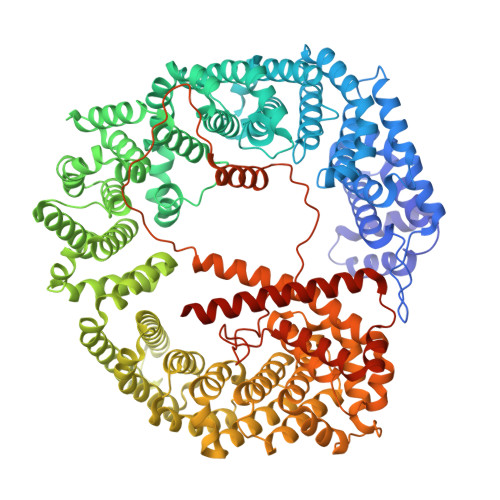
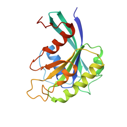
Structure of Importin 7 in complex with RanGTP


Experimental Data Snapshot

  • Method: ELECTRON MICROSCOPY
  • Resolution: 3.60 Å
  • Aggregation State: PARTICLE 
  • Reconstruction Method: SINGLE PARTICLE 

wwPDB Validation 3D Report Full Report

Validation slider image for 30JZ

This is version 1.1 of the entry. See complete history. 

Literature

Chaperoning by dynamic encasement: Structural basis of linker histone H1 nuclear import.

Fu, Z., Chafra, F., Freytag, B., Huyton, T., Gorlich, D.

(2026) Structure 

  • DOI: https://doi.org/10.1016/j.str.2026.09.001
  • Primary Citation Related Structures: 
    30FM, 30HD, 30JZ

  • PubMed Abstract: 

    Importins transport numerous proteins into cell nuclei. They also chaperone highly positively charged cargoes against ionic aggregation during transit. An extreme example is histone H1, which requires two importins, Importin 7 (Imp7) and Importin β (Impβ), for safe import. Here, we present a combined cryoelectron microscopy (cryo-EM)/molecular dynamics (MD) structure of the Imp7·Impβ·H1 complex, correcting a previous model (PDB 6N88) with misassigned importin chains. Our structure reveals that Imp7 contacts Impβ via three interfaces and that this heterodimerization creates a large negatively charged cavity. The globular H1-domain is recognized by Imp7 alone. The highly cationic C-terminal H1-tail participates in fluid-like transient interactions, contacting the inner surface of Impβ before returning to the α-solenoid and long acidic loop of Imp7. This architecture and dynamics explain how the importins shield the histone's enormous positive charge while avoiding interactions too strong for efficient intranuclear cargo release. Finally, we report a RanGTP·Imp7 structure, illustrating how RanGTP dissociates the import complex inside nuclei.


  • Organizational Affiliation: 
    • Department of Cellular Logistics, Max Planck Institute for Multidisciplinary Sciences, am Fassberg 11, 37077 Göttingen, Germany.

Macromolecule Content 

  • Total Structure Weight: 140.24 kDa 
  • Atom Count: 9,648 
  • Modeled Residue Count: 1,186 
  • Deposited Residue Count: 1,213 
  • Unique protein chains: 2

Macromolecules

Find similar proteins by:|  3D Structure
Entity ID: 1
MoleculeChains  Sequence LengthOrganismDetailsImage
Importin 7 L homeolog1,038Xenopus laevisMutation(s): 0 
Gene Names: ipo7.L, imp7, ipo7, MGC52556, ranbp7
UniProt
Find proteins for O42480 (Xenopus laevis)
Explore O42480 
Go to UniProtKB:  O42480
Entity Groups
Sequence Clusters30% Identity50% Identity70% Identity90% Identity95% Identity100% Identity
UniProt GroupO42480
Sequence Annotations
Expand
Reference Sequence
Find similar proteins by:|  3D Structure
Entity ID: 2
MoleculeChains  Sequence LengthOrganismDetailsImage
GTP-binding nuclear protein Ran175Homo sapiensMutation(s): 0 
Gene Names: RAN, ARA24, OK/SW-cl.81
EC: 3.6.5
UniProt & NIH Common Fund Data Resources
Find proteins for P62826 (Homo sapiens)
Explore P62826 
Go to UniProtKB:  P62826
PHAROS:  P62826
GTEx:  ENSG00000132341 
Entity Groups
Sequence Clusters30% Identity50% Identity70% Identity90% Identity95% Identity100% Identity
UniProt GroupP62826
Sequence Annotations
Expand
Reference Sequence

Experimental Data & Validation

Experimental Data

  • Method: ELECTRON MICROSCOPY
  • Resolution: 3.60 Å
  • Aggregation State: PARTICLE 
  • Reconstruction Method: SINGLE PARTICLE 
EM Software:
TaskSoftware PackageVersion
RECONSTRUCTIONRELION5.0.1
MODEL REFINEMENTPHENIX2.0_5885

Structure Validation

View Full Validation Report


Entry History 

& Funding Information

Deposition Data


Funding OrganizationLocationGrant Number
Max Planck SocietyGermany--

Revision History  (Full details and data files)

  • Version 1.0: 2026-09-23
    Type: Initial release
  • Version 1.1: 2026-10-07
    Changes: Data collection, Database references