Download Mendeley
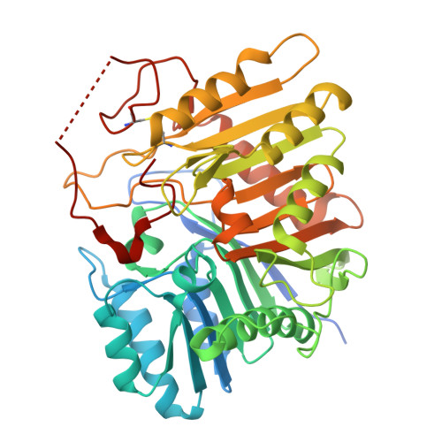
High-resolution Structure of the Vaccinia Virus Phospholipase D-fold Endonuclease K4.
Groger, H., Trouba, C., Barbaste, J., Lacroix, G., Schoehn, G., Burmeister, W.P., Tarbouriech, N.(2026) J Mol Biol 438: 169961-169961
- PubMed: 42526583 Search on PubMed
- DOI: https://doi.org/10.1016/j.jmb.2026.169961
- Primary Citation Related Structures:
30IE, 30MN - PubMed Abstract:
Vaccinia virus (VACV) is an orthopoxvirus closely related to mpox virus, which started global outbreaks in 2022. Poxvirus genomes are flanked by short, inverted complementary hairpin telomeres that feature mismatched bases and insertions essential for viral replication. In this context, a role of the late protein K4 has been proposed. K4 is present in the virion, is apparently non-essential and has a phospholipase D (PLD)-fold as has VACV F13 protein. It also shares fold and nuclease activity with its closest homologue, mammalian PLD3. We established an endonuclease activity against ssDNA and hairpin loops and bubbles in a dsDNA context while RNA is resistant to cleavage. The 2.4 Å cryo-EM structure of K4 shows an unusual octameric assembly, also present in solution. At low concentration, tetramers and dimers similar to the one of hPLD3 are also present. Despite its nuclease activity, in K4 a C-terminal extension blocks the DNA binding pockets. Using an inactive mutant, fortuitously, a DNA 19mer bound simultaneously to 2 sites of the octamer where it displaced the C-termini. DNA binding uses similar residues as the hPLD3 5'-exonuclease, despite different activities and orientations of the DNA. The role of K4 and the control of its activity by the observed auto-inhibition remain enigmatic.
- Univ. Grenoble Alpes, CEA, CNRS, IBS, F-38000 Grenoble, France.
Organizational Affiliation: