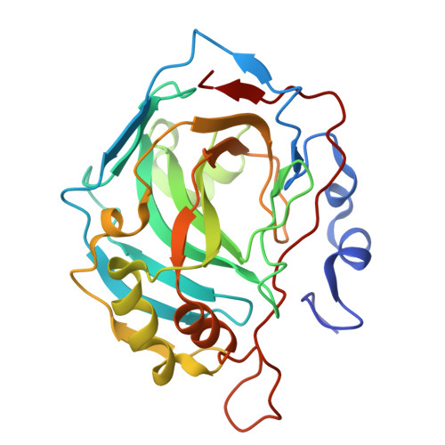
30IC | pdb_000030ic

Crystal structure of carbonic anhydrase II with methyl 4-methylsulfinyl-3-sulfamoyl-benzoate


Experimental Data Snapshot

  • Method: X-RAY DIFFRACTION
  • Resolution: 1.41 Å
  • R-Value Free: 
    0.203 (Depositor), 0.202 (DCC) 
  • R-Value Work: 
    0.169 (Depositor), 0.169 (DCC) 
  • R-Value Observed: 
    0.172 (Depositor) 

Starting Model: experimental
View more details

wwPDB Validation 3D Report Full Report

Validation slider image for 30IC

Ligand Structure Quality Assessment 


This is version 1.0 of the entry. See complete history. 

Literature

Enthalpy-Entropy Compensation in Inhibitor Binding to Carbonic Anhydrase Isozymes.

Paketuryte-Latve, V., Zaksauskas, A., Kairys, V., Capkauskaite, E., Bagdonas, M., Smirnov, A., Manakova, E., Grazulis, S., Matulis, D.

(2026) Biophys J 

  • DOI: https://doi.org/10.1016/j.bpj.2026.09.032
  • Primary Citation Related Structures: 
    30EN, 30EO, 30FA, 30FB, 30FN, 30FP, 30IC

  • PubMed Abstract: 

    Enthalpy-entropy compensation is a widely observed feature of protein-ligand recognition, yet its structural origins remain difficult to resolve. Here, we investigate a matched series of sulfonamide inhibitors binding to three closely related human carbonic anhydrase (CA) isozymes (CA II, CA XII, and CA XIII) using isothermal titration calorimetry, fluorescence thermal shift assays, and high-resolution X-ray crystallography. Although these isozymes share nearly identical overall folds and exhibit highly similar intrinsic Gibbs energies of binding, their enthalpic and entropic contributions diverge markedly. Correction of all observed thermodynamic quantities for proton-linked equilibria reveals that this divergence originates from subtle differences in the active-site microenvironment. In particular, single-residue variations-including the Thr/Thr/Val substitution at position 200, the Phe/Ala/Phe variation at position 131, and the Asn/Asn/Ser difference at position 62-modulate hydrogen-bond geometry, steric complementarity, and local solvation, thereby reshaping the balance between enthalpic and entropic driving forces. These results demonstrate how closely related proteins can achieve similar binding affinities through distinct energetic strategies and establish a coherent structure-thermodynamics framework for sulfonamide recognition.


  • Organizational Affiliation: 
    • Department of Biothermodynamics and Drug Design, Institute of Biotechnology, Life Sciences Center, Vilnius University, Vilnius, Lithuania.

Macromolecule Content 

  • Total Structure Weight: 30.15 kDa 
  • Atom Count: 2,520 
  • Modeled Residue Count: 258 
  • Deposited Residue Count: 260 
  • Unique protein chains: 1

Macromolecules

Find similar proteins by:|  3D Structure
Entity ID: 1
MoleculeChains  Sequence LengthOrganismDetailsImage
Carbonic anhydrase 2260Homo sapiensMutation(s): 0 
Gene Names: CA2
EC: 4.2.1.1 (PDB Primary Data), 4.2.1.69 (UniProt)
UniProt & NIH Common Fund Data Resources
Find proteins for P00918 (Homo sapiens)
Explore P00918 
Go to UniProtKB:  P00918
PHAROS:  P00918
GTEx:  ENSG00000104267 
Entity Groups
Sequence Clusters30% Identity50% Identity70% Identity90% Identity95% Identity100% Identity
UniProt GroupP00918
Sequence Annotations
Expand
Reference Sequence

Small Molecules

Ligands 5 Unique
IDChains Name / Formula / InChI Key2D Diagram3D Interactions
A1J55(
Subject of Investigation/LOI)

Query on A1J55


Download:Ideal Coordinates CCD File
E [auth A]methyl 4-methylsulfinyl-3-sulfamoyl-benzoate
C9 H11 N O5 S2
VYOFEEYRTWDOBI-MRXNPFEDSA-N
A1J56(
Subject of Investigation/LOI)

Query on A1J56


Download:Ideal Coordinates CCD File
F [auth A]methyl 4-methylsulfinyl-3-sulfamoyl-benzoate
C9 H11 N O5 S2
VYOFEEYRTWDOBI-INIZCTEOSA-N
BCN

Query on BCN


Download:Ideal Coordinates CCD File
D [auth A]BICINE
C6 H13 N O4
FSVCELGFZIQNCK-UHFFFAOYSA-N
DMS

Query on DMS


Download:Ideal Coordinates CCD File
C [auth A]DIMETHYL SULFOXIDE
C2 H6 O S
IAZDPXIOMUYVGZ-UHFFFAOYSA-N
ZN

Query on ZN


Download:Ideal Coordinates CCD File
B [auth A]ZINC ION
Zn
PTFCDOFLOPIGGS-UHFFFAOYSA-N

Experimental Data & Validation

Experimental Data

  • Method: X-RAY DIFFRACTION
  • Resolution: 1.41 Å
  • R-Value Free:  0.203 (Depositor), 0.202 (DCC) 
  • R-Value Work:  0.169 (Depositor), 0.169 (DCC) 
  • R-Value Observed: 0.172 (Depositor) 
Space Group: P 1 21 1
Unit Cell:
Length ( Å )Angle ( ˚ )
a = 42.209α = 90
b = 41.057β = 104.31
c = 71.909γ = 90
Software Package:
Software NamePurpose
REFMACrefinement
SCALAdata scaling
PDB_EXTRACTdata extraction
XDSdata reduction
MOLREPphasing

Structure Validation

View Full Validation Report


Ligand Structure Quality Assessment 


Entry History 

& Funding Information

Deposition Data


Funding OrganizationLocationGrant Number
Not funded--

Revision History  (Full details and data files)

  • Version 1.0: 2026-10-21
    Type: Initial release