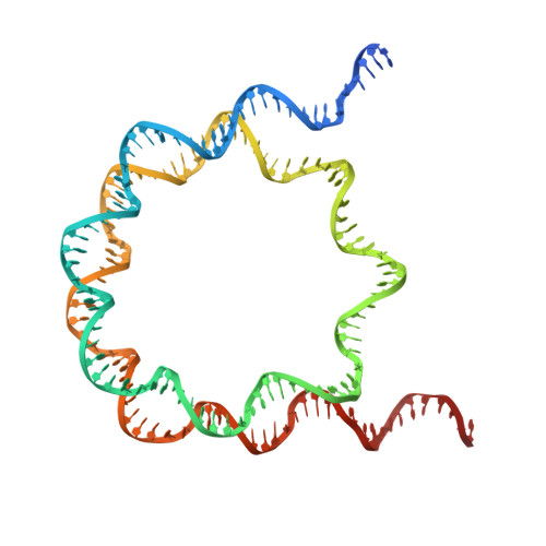
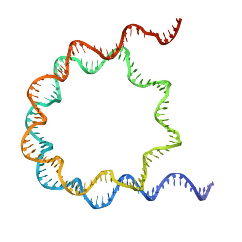
30GF | pdb_000030gf

Structural characterisation of chromatin remodelling intermediates supports linker DNA dependent product inhibition as a mechanism for nucleosome spacing.


Experimental Data Snapshot

  • Method: ELECTRON MICROSCOPY
  • Resolution: 4.80 Å
  • Aggregation State: PARTICLE 
  • Reconstruction Method: SINGLE PARTICLE 

Starting Models: experimental
View more details

wwPDB Validation 3D Report Full Report

Validation slider image for 30GF

This is version 1.0 of the entry. See complete history

Literature

Structural characterisation of chromatin remodelling intermediates supports linker DNA-dependent product inhibition as a mechanism for nucleosome spacing.

Hughes, A.L.Sundaramoorthy, R.Owen-Hughes, T.

(2025) Elife 14

  • DOI: https://doi.org/10.7554/eLife.52513
  • Primary Citation Related Structures: 
    30GF, 9R5K, 9R5S, 9R5W

  • PubMed Abstract: 

    Previously we showed that Saccharomyces cerevisiae Chd1 chromatin remodelling enzyme associates with nucleosomes oriented towards the longer linker (Sundaramoorthy et al., 2018) (1). Here we report a series of structures of Chd1 bound to nucleosomes during ongoing ATP-dependent repositioning. Combining these with biochemical experiments and existing literature we propose a model in which Chd1 first associates oriented to sample putative entry DNA. In an ATP-dependent reaction, the enzyme then redistributes to the opposite side of the nucleosome, where it subsequently adopts a conformation productive for DNA translocation. Once this active complex extends nascent exit linker to approximately 15bp, it is sensed by the Chd1 DNA binding domain resulting in conversion to a product inhibited state. These observations provide a mechanistic basis for the action of a molecular ruler element in nucleosome spacing.


  • Organizational Affiliation
    • Molecular Cell and Developmental Biology, University of Dundee, Dundee, United Kingdom.

Macromolecule Content 

  • Total Structure Weight: 210.15 kDa 
  • Atom Count: 12,169 
  • Modeled Residue Count: 1,050 
  • Deposited Residue Count: 1,316 
  • Unique protein chains: 4
  • Unique nucleic acid chains: 2

Macromolecules


Find similar proteins by:|  3D Structure
Entity ID: 1
MoleculeChains  Sequence LengthOrganismDetailsImage
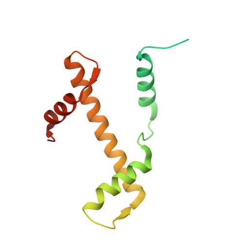
Histone H3.2
A, E
136Saccharomyces cerevisiaeMutation(s): 0 
Entity Groups
Sequence Clusters30% Identity50% Identity70% Identity90% Identity95% Identity100% Identity
Sequence Annotations
Expand
Reference Sequence
Find similar proteins by:|  3D Structure
Entity ID: 2
MoleculeChains  Sequence LengthOrganismDetailsImage
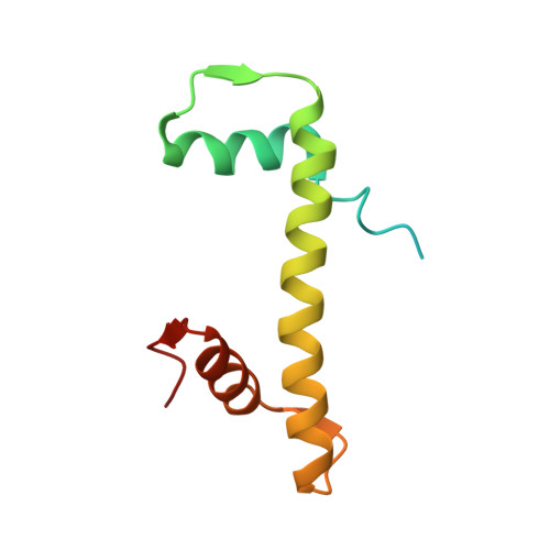
Histone H4
B, F
103Saccharomyces cerevisiaeMutation(s): 0 
Entity Groups
Sequence Clusters30% Identity50% Identity70% Identity90% Identity95% Identity100% Identity
Sequence Annotations
Expand
Reference Sequence
Find similar proteins by:|  3D Structure
Entity ID: 3
MoleculeChains  Sequence LengthOrganismDetailsImage
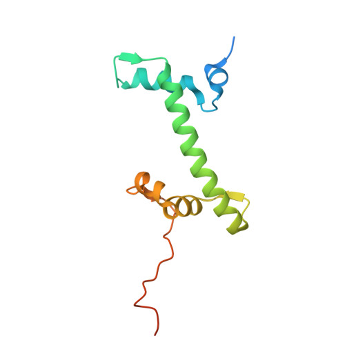
Histone H2A
C, G
130Saccharomyces cerevisiaeMutation(s): 0 
Entity Groups
Sequence Clusters30% Identity50% Identity70% Identity90% Identity95% Identity100% Identity
Sequence Annotations
Expand
Reference Sequence
Find similar proteins by:|  3D Structure
Entity ID: 4
MoleculeChains  Sequence LengthOrganismDetailsImage
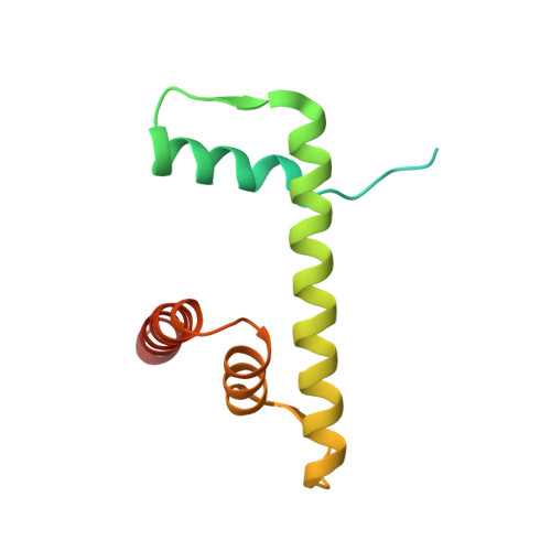
Histone H2B
D, H
126Saccharomyces cerevisiaeMutation(s): 0 
Entity Groups
Sequence Clusters30% Identity50% Identity70% Identity90% Identity95% Identity100% Identity
Sequence Annotations
Expand
Reference Sequence
Find similar nucleic acids by:  (by identity cutoff) 
Entity ID: 5
MoleculeChains LengthOrganismImage
DNA (154-MER)163synthetic construct
Sequence Annotations
Expand
Reference Sequence
Find similar nucleic acids by:  (by identity cutoff) 
Entity ID: 6
MoleculeChains LengthOrganismImage
DNA (154-MER)163synthetic construct
Sequence Annotations
Expand
Reference Sequence

Experimental Data & Validation

Experimental Data

  • Method: ELECTRON MICROSCOPY
  • Resolution: 4.80 Å
  • Aggregation State: PARTICLE 
  • Reconstruction Method: SINGLE PARTICLE 
EM Software:
TaskSoftware PackageVersion
RECONSTRUCTIONcryoSPARC4.5.3
MODEL REFINEMENTPHENIX1.21_5207

Structure Validation

View Full Validation Report


Entry History 

& Funding Information

Deposition Data


Funding OrganizationLocationGrant Number
Medical Research Council (MRC, United Kingdom)United Kingdom--

Revision History  (Full details and data files)

  • Version 1.0: 2026-08-26
    Type: Initial release