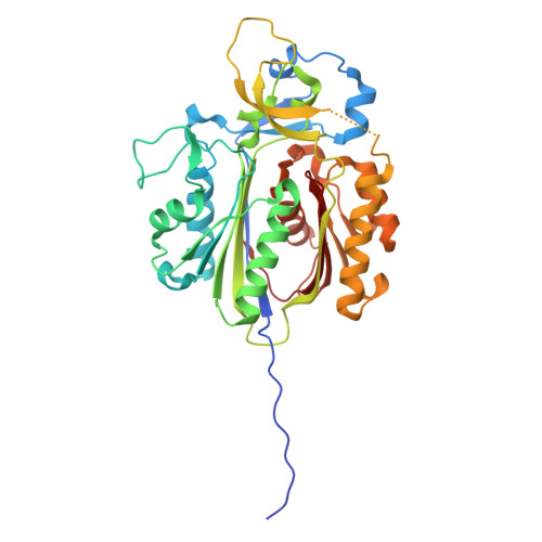
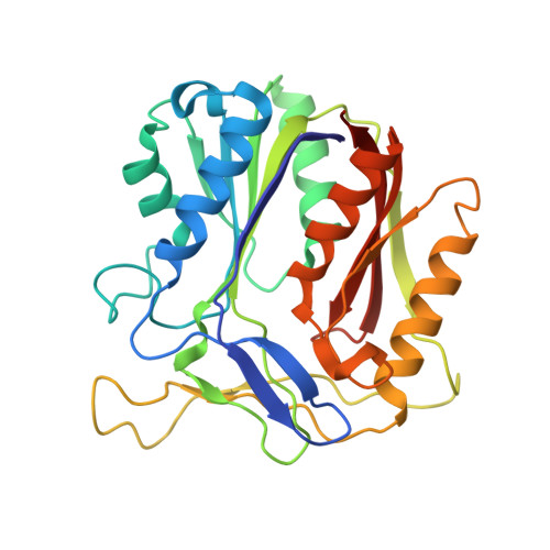
30EJ | pdb_000030ej

2-heptyl-4(1H)-quinolone synthase PqsBC in complex with compound 33


Experimental Data Snapshot

  • Method: X-RAY DIFFRACTION
  • Resolution: 2.06 Å
  • R-Value Free: 
    0.236 (Depositor), 0.232 (DCC) 
  • R-Value Work: 
    0.201 (Depositor), 0.199 (DCC) 
  • R-Value Observed: 
    0.203 (Depositor) 

Starting Model: experimental
View more details

wwPDB Validation 3D Report Full Report

Validation slider image for 30EJ

Ligand Structure Quality Assessment 


This is version 1.0 of the entry. See complete history. 

Literature

Chemical Inhibition of Pyocyanin Biosynthesis Attenuates Virulence of Pseudomonas aeruginosa.

Ding, W., Borgert, S.R., Blankenfeldt, W., Sieber, S.A.

(2026) Angew Chem Int Ed Engl 

  • DOI: https://doi.org/10.1002/anie.1294720
  • Primary Citation Related Structures: 
    30EG, 30EH, 30EJ

  • PubMed Abstract: 

    Anti-virulence strategies that disarm bacterial pathogenicity without affecting viability offer a promising alternative to traditional antibiotics by reducing selective pressure for resistance. However, in Gram-negative bacteria such as Pseudomonas aeruginosa, the double-layered cell envelope limits intracellular access of small-molecule inhibitors. Here, we screened a structurally diverse library of virulence inhibitors, previously established as pathoblockers in Gram-positive bacteria, and identified a potent β-lactone inhibitor of pyocyanin production. Activity-based protein profiling revealed PqsC, an intracellular enzyme involved in Pseudomonas quinolone signal (PQS) quorum sensing, as the primary molecular target. Accordingly, global proteomic analysis revealed widespread dysregulation of enzymes associated with the pyocyanin biosynthetic pathway. Biochemical assays confirmed inhibition of recombinant PqsBC, while intact protein mass spectrometry and site-directed mutagenesis established active site Cys129 as the covalent binding site. X-ray crystallography of the inhibited complex revealed a covalent binding mode. Subsequent virtual screening identified a chloroacetamide scaffold that exhibited enhanced potency and a well-defined binding mode. Importantly, supernatants from treated bacterial cultures showed reduced cytotoxicity toward mammalian cells, consistent with attenuated virulence. Collectively, this study identifies novel covalent inhibitors of PqsC and provides a mechanistic framework for the development of pathoblockers against P. aeruginosa.


  • Organizational Affiliation: 
    • Center for Functional Protein Assemblies (CPA), Department of Bioscience, TUM School of Natural Sciences, Technical University of Munich (TUM), Garching, Germany.

Macromolecule Content 

  • Total Structure Weight: 284.86 kDa 
  • Atom Count: 19,912 
  • Modeled Residue Count: 2,512 
  • Deposited Residue Count: 2,592 
  • Unique protein chains: 2

Macromolecules

Find similar proteins by:|  3D Structure
Entity ID: 1
MoleculeChains  Sequence LengthOrganismDetailsImage
2-heptyl-4(1H)-quinolone synthase subunit PqsC
A, C, E, G
365Pseudomonas aeruginosa PAO1Mutation(s): 0 
Gene Names: pqsC, PA0998
EC: 2.3.1.230
UniProt
Find proteins for Q9I4X1 (Pseudomonas aeruginosa (strain ATCC 15692 / DSM 22644 / CIP 104116 / JCM 14847 / LMG 12228 / 1C / PRS 101 / PAO1))
Explore Q9I4X1 
Go to UniProtKB:  Q9I4X1
Entity Groups
Sequence Clusters30% Identity50% Identity70% Identity90% Identity95% Identity100% Identity
UniProt GroupQ9I4X1
Sequence Annotations
Expand
Reference Sequence
Find similar proteins by:|  3D Structure
Entity ID: 2
MoleculeChains  Sequence LengthOrganismDetailsImage
2-heptyl-4(1H)-quinolone synthase subunit PqsB
B, D, F, H
283Pseudomonas aeruginosa PAO1Mutation(s): 0 
Gene Names: pqsB, PA0997
UniProt
Find proteins for Q9I4X2 (Pseudomonas aeruginosa (strain ATCC 15692 / DSM 22644 / CIP 104116 / JCM 14847 / LMG 12228 / 1C / PRS 101 / PAO1))
Explore Q9I4X2 
Go to UniProtKB:  Q9I4X2
Entity Groups
Sequence Clusters30% Identity50% Identity70% Identity90% Identity95% Identity100% Identity
UniProt GroupQ9I4X2
Sequence Annotations
Expand
Reference Sequence

Small Molecules

Ligands 3 Unique
IDChains Name / Formula / InChI Key2D Diagram3D Interactions
A1J5V
(Subject of Investigation/LOI)

Query on A1J5V


Download:Ideal Coordinates CCD File
J [auth A],
M [auth C],
N [auth E],
O [auth G]
N-[4-[3,4-bis(fluoranyl)phenyl]-1,3-thiazol-2-yl]-2-chloranyl-ethanamide
C11 H7 Cl F2 N2 O S
RHYUQCLMSGAKMN-UHFFFAOYSA-N
EDO

Query on EDO


Download:Ideal Coordinates CCD File
I [auth A],
L [auth B]
1,2-ETHANEDIOL
C2 H6 O2
LYCAIKOWRPUZTN-UHFFFAOYSA-N
CL

Query on CL


Download:Ideal Coordinates CCD File
K [auth A]CHLORIDE ION
Cl
VEXZGXHMUGYJMC-UHFFFAOYSA-M

Experimental Data & Validation

Experimental Data

  • Method: X-RAY DIFFRACTION
  • Resolution: 2.06 Å
  • R-Value Free:  0.236 (Depositor), 0.232 (DCC) 
  • R-Value Work:  0.201 (Depositor), 0.199 (DCC) 
  • R-Value Observed: 0.203 (Depositor) 
Space Group: C 1 2 1
Unit Cell:
Length ( Å )Angle ( ˚ )
a = 158.217α = 90
b = 226.862β = 112.33
c = 134.572γ = 90
Software Package:
Software NamePurpose
PHENIXrefinement
PDB_EXTRACTdata extraction
autoPROCdata processing
STARANISOdata scaling
PHASERphasing
XDSdata reduction

Structure Validation

View Full Validation Report


Ligand Structure Quality Assessment 


Entry History 

& Funding Information

Deposition Data


Funding OrganizationLocationGrant Number
German Research Foundation (DFG)Germany--

Revision History  (Full details and data files)

  • Version 1.0: 2026-11-11
    Type: Initial release