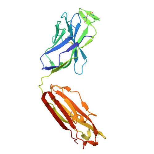
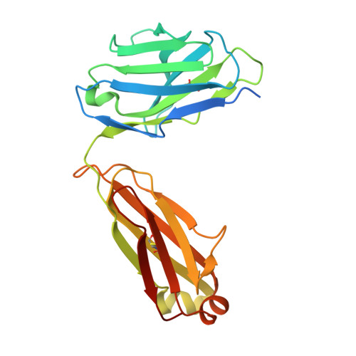
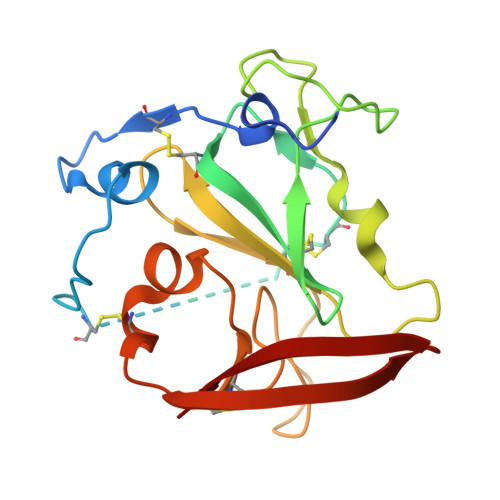
30CP | pdb_000030cp

Structure of the Hepatitis C Virus E2 Core from Genotype 6a in Complex with Germline Reverted Variants of the Broadly Neutralizing Antibody AR3C


Experimental Data Snapshot

  • Method: X-RAY DIFFRACTION
  • Resolution: 1.93 Å
  • R-Value Free: 
    0.245 (Depositor), 0.250 (DCC) 
  • R-Value Work: 
    0.202 (Depositor), 0.206 (DCC) 
  • R-Value Observed: 
    0.204 (Depositor) 

Starting Model: experimental
View more details

wwPDB Validation 3D Report Full Report

Validation slider image for 30CP

Ligand Structure Quality Assessment 


This is version 1.1 of the entry. See complete history. 

Literature

Structural and biochemical insights into HCV envelope proteins for germline-targeting vaccines.

Yechezkel, I., Maimon, H.E., Tennenhouse, A., Chen, F., Tarabih, H., Weisz, J., Fraenkel, R., Fleishman, S.J., Law, M., Tzarum, N.

(2026) PLoS Pathog 22: e1014651-e1014651

  • DOI: https://doi.org/10.1371/journal.ppat.1014651
  • Primary Citation Related Structures: 
    30BC, 30BG, 30BH, 30CP

  • PubMed Abstract: 

    Despite effective antivirals, Hepatitis C remains a global health burden, reflecting the difficulty of eliciting broadly neutralizing antibodies (bnAbs). For the Hepatitis C virus, bnAb responses are biased toward VH1-69-encoded Abs targeting a conserved epitope on the E2 glycoprotein, suggesting that effective immunogens must engage the corresponding germline (GL) B cell receptors. However, the structural basis of GL recognition remains unclear. Here, we show that GL-reverted VH1-69 bnAbs recognize the E2 core domain in a manner closely resembling mature Abs binding. High-resolution structures reveal a conserved mode of engagement, indicating that key epitope features are accessible prior to affinity maturation. Guided by these insights, we engineered E2 variants to enhance GL Ab interactions and stability. Although these designs increased affinity, they did not improve engagement of naïve B cells, highlighting a key constraint in GL-targeting strategies. Together, our findings define principles governing GL recognition and inform the rational design of an HCV vaccine.


  • Organizational Affiliation: 
    • Department of Biological Chemistry, Alexander Silberman Institute of Life Sciences, Faculty of Mathematics & Science, The Hebrew University of Jerusalem, Jerusalem, Israel.

Macromolecule Content 

  • Total Structure Weight: 72.47 kDa 
  • Atom Count: 5,101 
  • Modeled Residue Count: 608 
  • Deposited Residue Count: 654 
  • Unique protein chains: 3

Macromolecules

Find similar proteins by:|  3D Structure
Entity ID: 1
MoleculeChains  Sequence LengthOrganismDetailsImage
Fab AR3C GL heavy chainA [auth H]232Homo sapiensMutation(s): 0 
Entity Groups
Sequence Clusters30% Identity50% Identity70% Identity90% Identity95% Identity100% Identity
Sequence Annotations
Expand
Reference Sequence
Find similar proteins by:|  3D Structure
Entity ID: 2
MoleculeChains  Sequence LengthOrganismDetailsImage
Fab AR3C GL light chainB [auth L]233Homo sapiensMutation(s): 0 
Entity Groups
Sequence Clusters30% Identity50% Identity70% Identity90% Identity95% Identity100% Identity
Sequence Annotations
Expand
Reference Sequence
Find similar proteins by:|  3D Structure
Entity ID: 3
MoleculeChains  Sequence LengthOrganismDetailsImage
HCV E2 envelope proteinC [auth D0A0]189Recombinant Hepatitis C virus HK6a/JFH-1Mutation(s): 0 
Entity Groups
Sequence Clusters30% Identity50% Identity70% Identity90% Identity95% Identity100% Identity
Glycosylation
Glycosylation Sites: 3
Sequence Annotations
Expand
Reference Sequence

Oligosaccharides

Help  
Entity ID: 4
MoleculeChains Length2D Diagram GlycosylationD Interactions
2-acetamido-2-deoxy-beta-D-glucopyranose-(1-4)-2-acetamido-2-deoxy-beta-D-glucopyranoseD [auth E]2N-Glycosylation
Glycosylation Resources
GlyTouCan: G42666HT
GlyCosmos: G42666HT
GlyGen: G42666HT

Small Molecules

Ligands 2 Unique
IDChains Name / Formula / InChI Key2D Diagram3D Interactions
NAG
(Subject of Investigation/LOI)

Query on NAG


Download:Ideal Coordinates CCD File
K [auth D0A0],
L [auth D0A0]
2-acetamido-2-deoxy-beta-D-glucopyranose
C8 H15 N O6
OVRNDRQMDRJTHS-FMDGEEDCSA-N
PEG

Query on PEG


Download:Ideal Coordinates CCD File
E [auth H]
F [auth H]
G [auth H]
H
I [auth H]
E [auth H],
F [auth H],
G [auth H],
H,
I [auth H],
J [auth L],
M [auth D0A0],
N [auth D0A0],
O [auth D0A0],
P [auth D0A0]
DI(HYDROXYETHYL)ETHER
C4 H10 O3
MTHSVFCYNBDYFN-UHFFFAOYSA-N

Experimental Data & Validation

Experimental Data

  • Method: X-RAY DIFFRACTION
  • Resolution: 1.93 Å
  • R-Value Free:  0.245 (Depositor), 0.250 (DCC) 
  • R-Value Work:  0.202 (Depositor), 0.206 (DCC) 
  • R-Value Observed: 0.204 (Depositor) 
Space Group: P 21 21 21
Unit Cell:
Length ( Å )Angle ( ˚ )
a = 60.334α = 90
b = 64.45β = 90
c = 183.32γ = 90
Software Package:
Software NamePurpose
PHASERphasing
PHENIXrefinement
EDNAdata reduction

Structure Validation

View Full Validation Report


Ligand Structure Quality Assessment 


Entry History 

& Funding Information

Deposition Data


Funding OrganizationLocationGrant Number
United States - Israel Binational Science Foundation (BSF)United States2021-165
Israel Science FoundationIsrael1600/21

Revision History  (Full details and data files)

  • Version 1.0: 2026-10-07
    Type: Initial release
  • Version 1.1: 2026-10-28
    Changes: Database references