2ZYC | pdb_00002zyc

Crystal structure of peptidoglycan hydrolase from Sphingomonas sp. A1


Experimental Data Snapshot

  • Method: X-RAY DIFFRACTION
  • Resolution: 1.74 Å
  • R-Value Free: 
    0.238 (Depositor), 0.236 (DCC) 
  • R-Value Work: 
    0.199 (Depositor), 0.199 (DCC) 
  • R-Value Observed: 
    0.201 (Depositor) 

wwPDB Validation 3D Report Full Report

Validation slider image for 2ZYC

This is version 1.3 of the entry. See complete history

Literature

Crystal structure of the glycosidase family 73 peptidoglycan hydrolase FlgJ

Hashimoto, W.Ochiai, A.Momma, K.Itoh, T.Mikami, B.Maruyama, Y.Murata, K.

(2009) Biochem Biophys Res Commun 381: 16-21

  • DOI: https://doi.org/10.1016/j.bbrc.2009.01.186
  • Primary Citation Related Structures: 
    2ZYC

  • PubMed Abstract: 

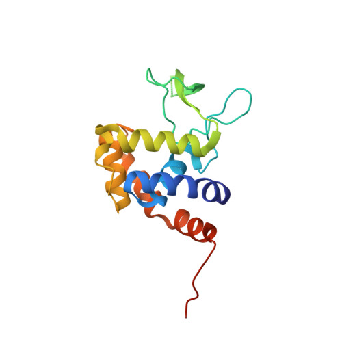
    Glycoside hydrolase (GH) categorized into family 73 plays an important role in degrading bacterial cell wall peptidoglycan. The flagellar protein FlgJ contains N- and C-terminal domains responsible for flagellar rod assembly and peptidoglycan hydrolysis, respectively. A member of family GH-73, the C-terminal domain (SPH1045-C) of FlgJ from Sphingomonas sp. strain A1 was expressed in Escherichia coli, purified, and characterized. SPH1045-C exhibited bacterial cell lytic activity most efficiently at pH 6.0 and 37 degrees C. The X-ray crystallographic structure of SPH1045-C was determined at 1.74 A resolution by single-wavelength anomalous diffraction. The enzyme consists of two lobes, alpha and beta. A deep cleft located between the two lobes can accommodate polymer molecules, suggesting that the active site is located in the cleft. Although SPH1045-C shows a structural homology with family GH-22 and GH-23 lysozymes, the arrangement of the nucleophile/base residue in the active site is specific to each peptidoglycan hydrolase.


  • Organizational Affiliation
    • Laboratory of Basic and Applied Molecular Biotechnology, Graduate School of Agriculture, Kyoto University, Uji, Kyoto 611-0011, Japan.

Macromolecule Content 

  • Total Structure Weight: 18.82 kDa 
  • Atom Count: 1,498 
  • Modeled Residue Count: 156 
  • Deposited Residue Count: 170 
  • Unique protein chains: 1

Macromolecules

Find similar proteins by:|  3D Structure
Entity ID: 1
MoleculeChains  Sequence LengthOrganismDetailsImage
Peptidoglycan hydrolase FlgJ170Sphingomonas sp. A1Mutation(s): 2 
Gene Names: flgJ
EC: 3.2.1
UniProt
Find proteins for B7XH69 (Sphingomonas sp. A1)
Explore B7XH69 
Go to UniProtKB:  B7XH69
Entity Groups
Sequence Clusters30% Identity50% Identity70% Identity90% Identity95% Identity100% Identity
UniProt GroupB7XH69
Sequence Annotations
Expand
Reference Sequence

Small Molecules

Ligands 1 Unique
IDChains Name / Formula / InChI Key2D Diagram3D Interactions
PO4

Query on PO4


Download:Ideal Coordinates CCD File
B [auth A]PHOSPHATE ION
O4 P
NBIIXXVUZAFLBC-UHFFFAOYSA-K

Experimental Data & Validation

Experimental Data

  • Method: X-RAY DIFFRACTION
  • Resolution: 1.74 Å
  • R-Value Free:  0.238 (Depositor), 0.236 (DCC) 
  • R-Value Work:  0.199 (Depositor), 0.199 (DCC) 
  • R-Value Observed: 0.201 (Depositor) 
Space Group: P 43 21 2
Unit Cell:
Length ( Å )Angle ( ˚ )
a = 54.28α = 90
b = 54.28β = 90
c = 102.75γ = 90
Software Package:
Software NamePurpose
REFMACrefinement
HKL-2000data collection
HKL-2000data reduction
HKL-2000data scaling
SHELXDEphasing

Structure Validation

View Full Validation Report


Entry History 

Deposition Data

Revision History  (Full details and data files)

  • Version 1.0: 2009-02-03
    Type: Initial release
  • Version 1.1: 2011-07-13
    Changes: Version format compliance
  • Version 1.2: 2021-11-10
    Changes: Database references, Derived calculations
  • Version 1.3: 2024-05-29
    Changes: Data collection