2ZXI | pdb_00002zxi

Structure of Aquifex aeolicus GidA in the form II crystal


Experimental Data Snapshot

  • Method: X-RAY DIFFRACTION
  • Resolution: 2.30 Å
  • R-Value Free: 
    0.248 (Depositor), 0.248 (DCC) 
  • R-Value Work: 
    0.209 (Depositor), 0.209 (DCC) 
  • R-Value Observed: 
    0.209 (Depositor) 

Starting Model: experimental
View more details

wwPDB Validation 3D Report Full Report

Validation slider image for 2ZXI

Ligand Structure Quality Assessment 


This is version 1.3 of the entry. See complete history

Literature

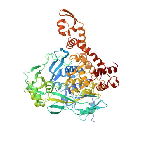
Conserved cysteine residues of GidA are essential for biogenesis of 5-carboxymethylaminomethyluridine at tRNA anticodon

Osawa, T.Ito, K.Inanaga, H.Nureki, O.Tomita, K.Numata, T.

(2009) Structure 17: 713-724

  • DOI: https://doi.org/10.1016/j.str.2009.03.013
  • Primary Citation Related Structures: 
    2ZXH, 2ZXI

  • PubMed Abstract: 

    The 5-carboxymethylaminomethyl modification of uridine (cmnm(5)U) at the anticodon first position occurs in tRNAs that read split codon boxes ending with purine. This modification is crucial for correct translation, by restricting codon-anticodon wobbling. Two conserved enzymes, GidA and MnmE, participate in the cmnm(5)U modification process. Here we determined the crystal structure of Aquifex aeolicus GidA at 2.3 A resolution. The structure revealed the tight interaction of GidA with FAD. Structure-based mutation analyses allowed us to identify two conserved Cys residues in the vicinity of the FAD-binding site that are essential for the cmnm(5)U modification in vivo. Together with mutational analysis of MnmE, we propose a mechanism for the cmnm(5)U modification process where GidA, but not MnmE, attacks the C6 atom of uridine by a mechanism analogous to that of thymidylate synthase. We also present a tRNA-docking model that provides structural insights into the tRNA recognition mechanism for efficient modification.


  • Organizational Affiliation
    • Institute for Biological Resources and Functions, National Institute of Advanced Industrial Science and Technology (AIST), Ibaraki, Japan.

Macromolecule Content 

  • Total Structure Weight: 290.44 kDa 
  • Atom Count: 20,580 
  • Modeled Residue Count: 2,415 
  • Deposited Residue Count: 2,548 
  • Unique protein chains: 1

Macromolecules

Find similar proteins by:|  3D Structure
Entity ID: 1
MoleculeChains  Sequence LengthOrganismDetailsImage
tRNA uridine 5-carboxymethylaminomethyl modification enzyme mnmG
A, B, C, D
637Aquifex aeolicusMutation(s): 0 
Gene Names: aq_761
UniProt
Find proteins for O66962 (Aquifex aeolicus (strain VF5))
Explore O66962 
Go to UniProtKB:  O66962
Entity Groups
Sequence Clusters30% Identity50% Identity70% Identity90% Identity95% Identity100% Identity
UniProt GroupO66962
Sequence Annotations
Expand
Reference Sequence

Experimental Data & Validation

Experimental Data

  • Method: X-RAY DIFFRACTION
  • Resolution: 2.30 Å
  • R-Value Free:  0.248 (Depositor), 0.248 (DCC) 
  • R-Value Work:  0.209 (Depositor), 0.209 (DCC) 
  • R-Value Observed: 0.209 (Depositor) 
Space Group: P 1 21 1
Unit Cell:
Length ( Å )Angle ( ˚ )
a = 119.414α = 90
b = 98.001β = 90.002
c = 129.622γ = 90
Software Package:
Software NamePurpose
CNSrefinement
HKL-2000data collection
HKL-2000data reduction
HKL-2000data scaling
PHASERphasing

Structure Validation

View Full Validation Report


Ligand Structure Quality Assessment 


Entry History 

Deposition Data

Revision History  (Full details and data files)

  • Version 1.0: 2009-05-19
    Type: Initial release
  • Version 1.1: 2011-07-13
    Changes: Version format compliance
  • Version 1.2: 2012-04-04
    Changes: Database references
  • Version 1.3: 2023-11-01
    Changes: Data collection, Database references, Derived calculations, Refinement description