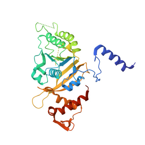
Functionally important movements in RecA molecules and filaments: studies involving mutation and environmental changes
Prabu, J.R., Manjunath, G.P., Chandra, N.R., Muniyappa, K., Vijayan, M.(2008) Acta Crystallogr D Biol Crystallogr 64: 1146-1157
- PubMed: 19020353
- DOI: https://doi.org/10.1107/S0907444908028448
- Primary Citation of Related Structures:
2ZR0, 2ZR7, 2ZR9, 2ZRA, 2ZRB, 2ZRC, 2ZRD, 2ZRE, 2ZRF, 2ZRG, 2ZRH, 2ZRI, 2ZRJ, 2ZRK, 2ZRL, 2ZRM, 2ZRN, 2ZRO, 2ZRP - PubMed Abstract:
The crystal structures of mutants of Mycobacterium smegmatis RecA (MsRecA) involving changes of Gln196 from glutamine to alanine, asparagine and glutamic acid, wild-type MsRecA and several of their nucleotide complexes have been determined using mostly low-temperature and partly room-temperature X-ray data. At both temperatures, nucleotide binding results in a movement of Gln196 towards the bound nucleotide in the wild-type protein. This movement is abolished in the mutants, thus establishing the structural basis for the triggering action of the residue in terms of the size, shape and the chemical nature of the side chain. The 19 crystal structures reported here, together with 11 previously reported MsRecA structures, provide further elaboration of the relation between the pitch of the ;inactive' RecA filament, the orientation of the C-terminal domain with respect to the main domain and the location of the switch residue. The low-temperature structures define one extreme of the range of positions the C-terminal domain can occupy. The movement of the C-terminal domain is correlated with those of the LexA-binding loop and the loop that connects the main and the N-terminal domains. These elements of molecular plasticity are made use of in the transition to the ;active' filament, as evidenced by the recently reported structures of RecA-DNA complexes. The available structures of RecA resulting from X-ray and electron-microscopic studies appear to represent different stages in the trajectory of the allosteric transformations of the RecA filament. The work reported here contributes to the description of the early stages of this trajectory and provides insight into structures relevant to the later stages.
- Molecular Biophysics Unit, Indian Institute of Science, Bangalore 560012, India.
Organizational Affiliation:


