2ZPP | pdb_00002zpp

Neutron crystal structure of cubic insulin at pD9


Experimental Data Snapshot

  • Method: NEUTRON DIFFRACTION
  • Resolution: 2.50 Å
  • R-Value Free: 
    0.260 (Depositor) 
  • R-Value Work: 
    0.221 (Depositor) 
  • R-Value Observed: 
    0.222 (Depositor) 

Starting Model: experimental
View more details

wwPDB Validation 3D Report Full Report

Validation slider image for 2ZPP

This is version 1.5 of the entry. See complete history

Literature

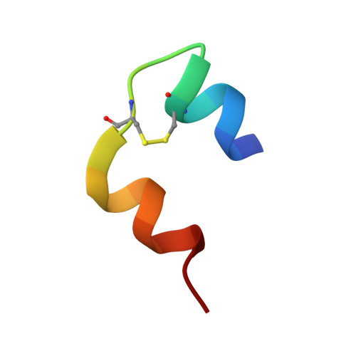
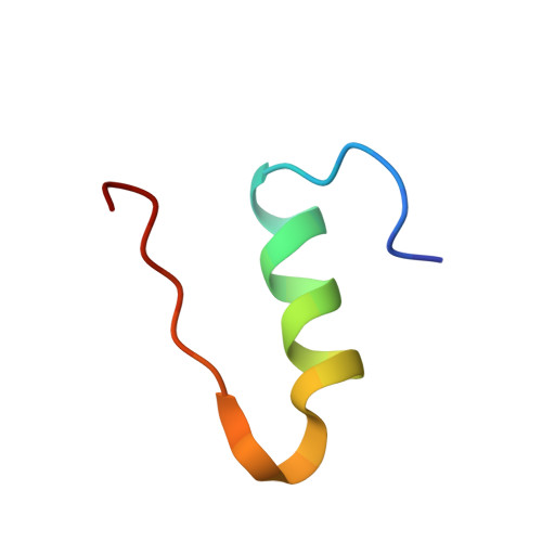
An abnormal pK(a) value of internal histidine of the insulin molecule revealed by neutron crystallographic analysis

Ishikawa, T.Chatake, T.Morimoto, Y.Maeda, M.Kurihara, K.Tanaka, I.Niimura, N.

(2008) Biochem Biophys Res Commun 376: 32-35

  • DOI: https://doi.org/10.1016/j.bbrc.2008.08.071
  • Primary Citation Related Structures: 
    2ZPP

  • PubMed Abstract: 

    Insulin is stored in pancreatic beta-cell as hexameric form with Zn2+ ions, while the hormonally active form is monomer. The hexamer requires the coordination of Zn2+ ions to the HisB10. In order to reveal the mechanism of the hexamerization of insulin, we investigated the Zn2+ free insulin at pD6.6 and pD9 by neutron crystallographic analyses. HisB10 is doubly protonated not only at pD6.6 but also at pD9, indicating an abnormal pK(a) of this histidine. It is suggested that HisB10 acts on a strong cation capture and contributes to the high stability of the hexameric form in pancreas.


  • Organizational Affiliation
    • Kyoto University Research Reactor, Asashironishi 2, Kumatori, Osaka 590-0494, Japan.

Macromolecule Content 

  • Total Structure Weight: 5.79 kDa 
  • Atom Count: 437 
  • Modeled Residue Count: 51 
  • Deposited Residue Count: 51 
  • Unique protein chains: 2

Macromolecules

Find similar proteins by:  Sequence   |   3D Structure  
Entity ID: 1
MoleculeChains  Sequence LengthOrganismDetailsImage
Insulin A chain21Sus scrofaMutation(s): 0 
UniProt
Find proteins for P01315 (Sus scrofa)
Explore P01315 
Go to UniProtKB:  P01315
Entity Groups
Sequence Clusters30% Identity50% Identity70% Identity90% Identity95% Identity100% Identity
UniProt GroupP01315
Sequence Annotations
Expand
Reference Sequence
Find similar proteins by:|  3D Structure
Entity ID: 2
MoleculeChains  Sequence LengthOrganismDetailsImage
Insulin B chain30Sus scrofaMutation(s): 0 
UniProt
Find proteins for P01315 (Sus scrofa)
Explore P01315 
Go to UniProtKB:  P01315
Entity Groups
Sequence Clusters30% Identity50% Identity70% Identity90% Identity95% Identity100% Identity
UniProt GroupP01315
Sequence Annotations
Expand
Reference Sequence

Experimental Data & Validation

Experimental Data

  • Method: NEUTRON DIFFRACTION
  • Resolution: 2.50 Å
  • R-Value Free:  0.260 (Depositor) 
  • R-Value Work:  0.221 (Depositor) 
  • R-Value Observed: 0.222 (Depositor) 
Space Group: I 21 3
Unit Cell:
Length ( Å )Angle ( ˚ )
a = 79.04α = 90
b = 79.04β = 90
c = 79.04γ = 90
Software Package:
Software NamePurpose
DENZOdata reduction
CNSrefinement
SCALEPACKdata scaling
CNSphasing

Structure Validation

View Full Validation Report


Entry History 

Deposition Data

Revision History  (Full details and data files)

  • Version 1.0: 2008-10-28
    Type: Initial release
  • Version 1.1: 2011-07-13
    Changes: Non-polymer description, Version format compliance
  • Version 1.2: 2013-08-21
    Changes: Other
  • Version 1.3: 2018-03-07
    Changes: Data collection
  • Version 1.4: 2023-11-01
    Changes: Data collection, Database references, Refinement description
  • Version 1.5: 2024-10-23
    Changes: Structure summary