2ZOK | pdb_00002zok

Crystal structure of H-2Db in complex with JHMV epitope S510


Experimental Data Snapshot

  • Method: X-RAY DIFFRACTION
  • Resolution: 2.10 Å
  • R-Value Free: 
    0.266 (Depositor), 0.260 (DCC) 
  • R-Value Work: 
    0.206 (Depositor), 0.202 (DCC) 
  • R-Value Observed: 
    0.209 (Depositor) 

Starting Model: experimental
View more details

wwPDB Validation 3D Report Full Report

Validation slider image for 2ZOK

This is version 1.4 of the entry. See complete history

Literature

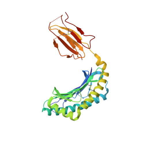
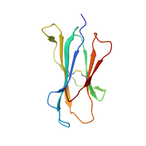
Structural and biological basis of CTL escape in coronavirus-infected mice.

Butler, N.S.Theodossis, A.Webb, A.I.Dunstone, M.A.Nastovska, R.Ramarathinam, S.H.Rossjohn, J.Purcell, A.W.Perlman, S.

(2008) J Immunol 180: 3926-3937

  • DOI: https://doi.org/10.4049/jimmunol.180.6.3926
  • Primary Citation Related Structures: 
    2ZOK, 2ZOL

  • PubMed Abstract: 

    Cytotoxic T lymphocyte escape occurs in many human infections, as well as mice infected with the JHM strain of mouse hepatitis virus, which exhibit CTL escape variants with mutations in a single epitope from the spike glycoprotein (S510). In all CTL epitopes prone to escape, only a subset of all potential variants is generally detected, even though many of the changes that are not selected would result in evasion of the T cell response. It is postulated that these unselected mutations significantly impair virus fitness. To define more precisely the basis for this preferential selection, we combine x-ray crystallographic studies of the MHC class I (D(b))/S510 complexes with viral reverse genetics to identify a prominent TCR contact residue (tryptophan at position 4) prone to escape mutations. The data show that a mutation that is commonly detected in chronically infected mice (tryptophan to arginine) potently disrupts the topology of the complex, explaining its selection. However, other mutations at this residue, which also abrogate the CTL response, are never selected in vivo even though they do not compromise virus fitness in acutely infected animals or induce a significant de novo CTL response. Thus, while structural analyses of the S510/D(b) complex provide a strong basis for why some CTL escape variants are selected, our results also show that factors other than effects on virus fitness limit the diversification of CD8 T cell epitopes.


  • Organizational Affiliation
    • Department of Microbiology, University of Iowa, Iowa City, IA 52242, USA.

Macromolecule Content 

  • Total Structure Weight: 181.98 kDa 
  • Atom Count: 13,084 
  • Modeled Residue Count: 1,458 
  • Deposited Residue Count: 1,548 
  • Unique protein chains: 3

Macromolecules

Find similar proteins by:|  3D Structure
Entity ID: 1
MoleculeChains  Sequence LengthOrganismDetailsImage
H-2 class I histocompatibility antigen, D-B alpha chain
A, C, E, G
278Mus musculusMutation(s): 0 
UniProt
Find proteins for P01899 (Mus musculus)
Explore P01899 
Go to UniProtKB:  P01899
Entity Groups
Sequence Clusters30% Identity50% Identity70% Identity90% Identity95% Identity100% Identity
UniProt GroupP01899
Sequence Annotations
Expand
Reference Sequence
Find similar proteins by:|  3D Structure
Entity ID: 2
MoleculeChains  Sequence LengthOrganismDetailsImage
Beta-2-microglobulin
B, D, F, H
100Mus musculusMutation(s): 0 
UniProt & NIH Common Fund Data Resources
Find proteins for P01887 (Mus musculus)
Explore P01887 
Go to UniProtKB:  P01887
IMPC:  MGI:88127
Entity Groups
Sequence Clusters30% Identity50% Identity70% Identity90% Identity95% Identity100% Identity
UniProt GroupP01887
Sequence Annotations
Expand
Reference Sequence
Find similar proteins by:  Sequence   |   3D Structure  
Entity ID: 3
MoleculeChains  Sequence LengthOrganismDetailsImage
9-meric peptide from Spike glycoproteinI,
J [auth L],
K [auth J],
L [auth K]
9N/AMutation(s): 1 
UniProt
Find proteins for Q02385 (Murine coronavirus (strain JHMV / variant CL-2))
Explore Q02385 
Go to UniProtKB:  Q02385
Entity Groups
UniProt GroupQ02385
Sequence Annotations
Expand
Reference Sequence

Small Molecules

Ligands 2 Unique
IDChains Name / Formula / InChI Key2D Diagram3D Interactions
SO4

Query on SO4


Download:Ideal Coordinates CCD File
M [auth A],
P [auth E],
R [auth G],
T [auth I]
SULFATE ION
O4 S
QAOWNCQODCNURD-UHFFFAOYSA-L
GOL

Query on GOL


Download:Ideal Coordinates CCD File
N [auth A],
O [auth C],
Q [auth E],
S [auth G]
GLYCEROL
C3 H8 O3
PEDCQBHIVMGVHV-UHFFFAOYSA-N
Modified Residues  1 Unique
IDChains TypeFormula2D DiagramParent
ABA
Query on ABA
I,
J [auth L],
K [auth J],
L [auth K]
L-PEPTIDE LINKINGC4 H9 N O2ALA

Experimental Data & Validation

Experimental Data

  • Method: X-RAY DIFFRACTION
  • Resolution: 2.10 Å
  • R-Value Free:  0.266 (Depositor), 0.260 (DCC) 
  • R-Value Work:  0.206 (Depositor), 0.202 (DCC) 
  • R-Value Observed: 0.209 (Depositor) 
Space Group: P 1 21 1
Unit Cell:
Length ( Å )Angle ( ˚ )
a = 79.538α = 90
b = 86.056β = 90.01
c = 152.072γ = 90
Software Package:
Software NamePurpose
REFMACrefinement
ADSCdata collection
MOSFLMdata reduction
SCALAdata scaling
PHASERphasing

Structure Validation

View Full Validation Report


Entry History 

Deposition Data

Revision History  (Full details and data files)

  • Version 1.0: 2008-06-10
    Type: Initial release
  • Version 1.1: 2011-07-13
    Changes: Non-polymer description, Version format compliance
  • Version 1.2: 2019-11-06
    Changes: Data collection, Database references, Derived calculations
  • Version 1.3: 2023-11-01
    Changes: Data collection, Database references, Derived calculations, Refinement description
  • Version 1.4: 2023-11-15
    Changes: Data collection