2ZMD | pdb_00002zmd

Crystal structure of human Mps1 catalytic domain T686A mutant in complex with SP600125 inhibitor


Experimental Data Snapshot

  • Method: X-RAY DIFFRACTION
  • Resolution: 2.88 Å
  • R-Value Free: 
    0.261 (Depositor) 
  • R-Value Work: 
    0.221 (Depositor) 
  • R-Value Observed: 
    0.223 (Depositor) 

Starting Model: experimental
View more details

wwPDB Validation 3D Report Full Report

Validation slider image for 2ZMD

Ligand Structure Quality Assessment 


This is version 1.3 of the entry. See complete history

Literature

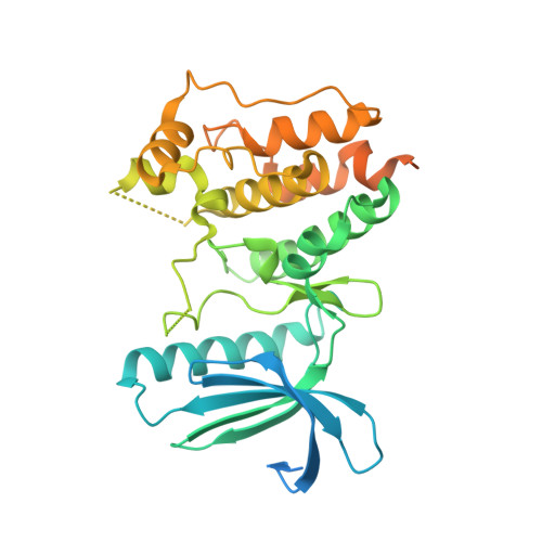
Crystal structure of the catalytic domain of the mitotic checkpoint kinase Mps1 in complex with SP600125.

Chu, M.L.Chavas, L.M.Douglas, K.T.Eyers, P.A.Tabernero, L.

(2008) J Biological Chem 283: 21495-21500

  • DOI: https://doi.org/10.1074/jbc.M803026200
  • Primary Citation Related Structures: 
    2ZMC, 2ZMD

  • PubMed Abstract: 

    Chromosomal instability can result from defective control of checkpoints and is associated with malignant cell growth. Monopolar spindle 1 (Mps1) is a dual-specificity protein kinase that has important roles in the prevention of aneuploidy during the cell cycle and might therefore be a potential target for new therapeutic agents in the treatment of cancer. To gain insights into the molecular mechanism of Mps1 inhibition by small molecules, we determined the x-ray structure of Mps1, both alone and in complex with the ATP-competitive inhibitor SP600125. Mps1 adopts a classic protein kinase fold, with the inhibitor sitting in the ATP-binding site where it is stabilized by hydrophobic interactions. We identified a secondary pocket, not utilized by SP600125, which might be exploited for the rational design of specific Mps1 inhibitors. These structures provide important insights into the interaction of this protein kinase with small molecules and suggest potential mechanisms for Mps1 regulation.


  • Organizational Affiliation
    • School of Pharmacy and Pharmaceutical Sciences, Stopford Building, University of Manchester, Manchester, UK.

Macromolecule Content 

  • Total Structure Weight: 44.85 kDa 
  • Atom Count: 2,170 
  • Modeled Residue Count: 259 
  • Deposited Residue Count: 390 
  • Unique protein chains: 1

Macromolecules

Find similar proteins by:|  3D Structure
Entity ID: 1
MoleculeChains  Sequence LengthOrganismDetailsImage
Dual specificity protein kinase TTK390Homo sapiensMutation(s): 1 
Gene Names: TTKMPS1L1
EC: 2.7.12.1
UniProt & NIH Common Fund Data Resources
Find proteins for P33981 (Homo sapiens)
Explore P33981 
Go to UniProtKB:  P33981
PHAROS:  P33981
GTEx:  ENSG00000112742 
Entity Groups
Sequence Clusters30% Identity50% Identity70% Identity90% Identity95% Identity100% Identity
UniProt GroupP33981
Sequence Annotations
Expand
Reference Sequence

Small Molecules

Ligands 2 Unique
IDChains Name / Formula / InChI Key2D Diagram3D Interactions
7PE

Query on 7PE


Download:Ideal Coordinates CCD File
C [auth A]2-(2-(2-(2-(2-(2-ETHOXYETHOXY)ETHOXY)ETHOXY)ETHOXY)ETHOXY)ETHANOL
C14 H30 O7
UKXKPKBTMYNOFS-UHFFFAOYSA-N
537

Query on 537


Download:Ideal Coordinates CCD File
B [auth A]2,6-DIHYDROANTHRA/1,9-CD/PYRAZOL-6-ONE
C14 H8 N2 O
ACPOUJIDANTYHO-UHFFFAOYSA-N

Experimental Data & Validation

Experimental Data

  • Method: X-RAY DIFFRACTION
  • Resolution: 2.88 Å
  • R-Value Free:  0.261 (Depositor) 
  • R-Value Work:  0.221 (Depositor) 
  • R-Value Observed: 0.223 (Depositor) 
Space Group: I 2 2 2
Unit Cell:
Length ( Å )Angle ( ˚ )
a = 70.6α = 90
b = 105.17β = 90
c = 111.97γ = 90
Software Package:
Software NamePurpose
REFMACrefinement
CrystalCleardata collection
MOSFLMdata reduction
SCALAdata scaling
MOLREPphasing

Structure Validation

View Full Validation Report


Ligand Structure Quality Assessment 


Entry History 

Deposition Data

Revision History  (Full details and data files)

  • Version 1.0: 2008-05-13
    Type: Initial release
  • Version 1.1: 2011-07-13
    Changes: Advisory, Version format compliance
  • Version 1.2: 2021-11-10
    Changes: Database references, Derived calculations
  • Version 1.3: 2023-11-01
    Changes: Data collection, Refinement description