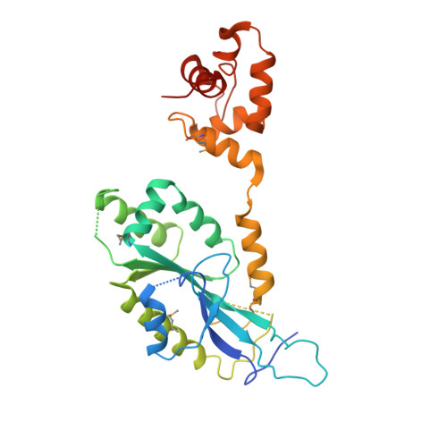
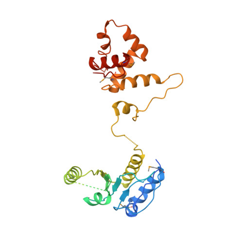
2ZIW

Crystal structure of the Mus81-Eme1 complex


Experimental Data Snapshot

  • Method: X-RAY DIFFRACTION
  • Resolution: 2.80 Å
  • R-Value Free: 0.327 
  • R-Value Work: 0.272 
  • R-Value Observed: 0.272 

wwPDB Validation   3D Report Full Report


This is version 1.2 of the entry. See complete history


Literature

Crystal structure of the Mus81-Eme1 complex

Chang, J.H.Kim, J.J.Choi, J.M.Lee, J.H.Cho, Y.

(2008) Genes Dev 22: 1093-1106

  • DOI: https://doi.org/10.1101/gad.1618708
  • Primary Citation of Related Structures:  
    2ZIU, 2ZIV, 2ZIW, 2ZIX

  • PubMed Abstract: 

    The Mus81-Eme1 complex is a structure-specific endonuclease that plays an important role in rescuing stalled replication forks and resolving the meiotic recombination intermediates in eukaryotes. We have determined the crystal structure of the Mus81-Eme1 complex. Both Mus81 and Eme1 consist of a central nuclease domain, two repeats of the helix-hairpin-helix (HhH) motif at their C-terminal region, and a linker helix. While each domain structure resembles archaeal XPF homologs, the overall structure is significantly different from those due to the structure of a linker helix. We show that a flexible intradomain linker that formed with 36 residues in the nuclease domain of Eme1 is essential for the recognition of DNA. We identified several basic residues lining the outer surface of the active site cleft of Mus81 that are involved in the interaction with a flexible arm of a nicked Holliday junction (HJ). These interactions might contribute to the optimal positioning of the opposite junction across the nick into the catalytic site, which provided the basis for the "nick and counternick" mechanism of Mus81-Eme1 and for the nicked HJ to be the favored in vitro substrate of this enzyme.


  • Organizational Affiliation

    National Creative Initiatives for Structural Biology and Department of Life Science, Pohang University of Science and Technology, Pohang, KyungBook 790-784, South Korea.


Macromolecules
Find similar proteins by:  (by identity cutoff)  |  3D Structure
Entity ID: 1
MoleculeChains Sequence LengthOrganismDetailsImage
Mus81 protein311Danio rerioMutation(s): 0 
Gene Names: mus81
EC: 3.1.22
UniProt
Find proteins for Q7SXA9 (Danio rerio)
Explore Q7SXA9 
Go to UniProtKB:  Q7SXA9
Entity Groups  
Sequence Clusters30% Identity50% Identity70% Identity90% Identity95% Identity100% Identity
UniProt GroupQ7SXA9
Sequence Annotations
Expand
  • Reference Sequence
Find similar proteins by:  (by identity cutoff)  |  3D Structure
Entity ID: 2
MoleculeChains Sequence LengthOrganismDetailsImage
Crossover junction endonuclease EME1341Homo sapiensMutation(s): 0 
Gene Names: EME1
EC: 3.1.22
UniProt & NIH Common Fund Data Resources
Find proteins for Q96AY2 (Homo sapiens)
Explore Q96AY2 
Go to UniProtKB:  Q96AY2
PHAROS:  Q96AY2
GTEx:  ENSG00000154920 
Entity Groups  
Sequence Clusters30% Identity50% Identity70% Identity90% Identity95% Identity100% Identity
UniProt GroupQ96AY2
Sequence Annotations
Expand
  • Reference Sequence
Small Molecules
Modified Residues  1 Unique
IDChains TypeFormula2D DiagramParent
MSE
Query on MSE
A
L-PEPTIDE LINKINGC5 H11 N O2 SeMET
Experimental Data & Validation

Experimental Data

  • Method: X-RAY DIFFRACTION
  • Resolution: 2.80 Å
  • R-Value Free: 0.327 
  • R-Value Work: 0.272 
  • R-Value Observed: 0.272 
  • Space Group: P 31 2 1
Unit Cell:
Length ( Å )Angle ( ˚ )
a = 88.963α = 90
b = 88.963β = 90
c = 169.326γ = 120
Software Package:
Software NamePurpose
CNSrefinement
HKL-2000data collection
HKL-2000data reduction
SCALEPACKdata scaling
SHELXSphasing

Structure Validation

View Full Validation Report


Entry History 

Deposition Data

Revision History  (Full details and data files)

  • Version 1.0: 2008-04-29
    Type: Initial release
  • Version 1.1: 2011-07-13
    Changes: Version format compliance
  • Version 1.2: 2024-10-30
    Changes: Data collection, Database references, Derived calculations, Structure summary