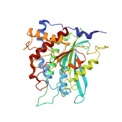
A conserved hydrogen-bond network in the catalytic centre of animal glutaminyl cyclases is critical for catalysis.
Huang, K.F., Wang, Y.R., Chang, E.C., Chou, T.L., Wang, A.H.(2008) Biochem J 411: 181-190
- PubMed: 18072935
- DOI: https://doi.org/10.1042/BJ20071073
- Primary Citation of Related Structures:
2ZED, 2ZEE, 2ZEF, 2ZEG, 2ZEH, 2ZEL, 2ZEM, 2ZEN, 2ZEO, 2ZEP - PubMed Abstract:
QCs (glutaminyl cyclases; glutaminyl-peptide cyclotransferases, EC 2.3.2.5) catalyse N-terminal pyroglutamate formation in numerous bioactive peptides and proteins. The enzymes were reported to be involved in several pathological conditions such as amyloidotic disease, osteoporosis, rheumatoid arthritis and melanoma. The crystal structure of human QC revealed an unusual H-bond (hydrogen-bond) network in the active site, formed by several highly conserved residues (Ser(160), Glu(201), Asp(248), Asp(305) and His(319)), within which Glu(201) and Asp(248) were found to bind to substrate. In the present study we combined steady-state enzyme kinetic and X-ray structural analyses of 11 single-mutation human QCs to investigate the roles of the H-bond network in catalysis. Our results showed that disrupting one or both of the central H-bonds, i.e., Glu(201)...Asp(305) and Asp(248)...Asp(305), reduced the steady-state catalysis dramatically. The roles of these two COOH...COOH bonds on catalysis could be partly replaced by COOH...water bonds, but not by COOH...CONH(2) bonds, reminiscent of the low-barrier Asp...Asp H-bond in the active site of pepsin-like aspartic peptidases. Mutations on Asp(305), a residue located at the centre of the H-bond network, raised the K(m) value of the enzyme by 4.4-19-fold, but decreased the k(cat) value by 79-2842-fold, indicating that Asp(305) primarily plays a catalytic role. In addition, results from mutational studies on Ser(160) and His(319) suggest that these two residues might help to stabilize the conformations of Asp(248) and Asp(305) respectively. These data allow us to propose an essential proton transfer between Glu(201), Asp(305) and Asp(248) during the catalysis by animal QCs.
- Institute of Biological Chemistry, Academia Sinica, Taipei 115, Taiwan.
Organizational Affiliation:


