2ZBZ | pdb_00002zbz

Crystal structure of vitamin D hydroxylase cytochrome P450 105A1 (R84A mutant) in complex with 1,25-dihydroxyvitamin D3


Experimental Data Snapshot

  • Method: X-RAY DIFFRACTION
  • Resolution: 1.90 Å
  • R-Value Free: 
    0.242 (Depositor), 0.240 (DCC) 
  • R-Value Work: 
    0.198 (Depositor), 0.200 (DCC) 
  • R-Value Observed: 
    0.200 (Depositor) 

Starting Model: experimental
View more details

wwPDB Validation   3D Report Full Report


Ligand Structure Quality Assessment 

Created with Raphaël 2.3.0Worse 01 BetterLigand structure goodness of fit to experimental dataBest fitted HEMClick on this verticalbar to view detailsBest fitted VDXClick on this verticalbar to view details

This is version 1.3 of the entry. See complete history


Literature

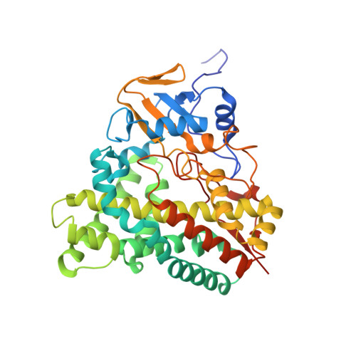
Crystal Structure of CYP105A1 (P450SU-1) in Complex with 1alpha,25-Dihydroxyvitamin D3

Sugimoto, H.Shinkyo, R.Hayashi, K.Yoneda, S.Yamada, M.Kamakura, M.Ikushiro, S.Shiro, Y.Sakaki, T.

(2008) Biochemistry 47: 4017-4027

  • DOI: https://doi.org/10.1021/bi7023767
  • Primary Citation of Related Structures:  
    2ZBX, 2ZBY, 2ZBZ

  • PubMed Abstract: 

    Vitamin D 3 (VD 3), a prohormone in mammals, plays a crucial role in the maintenance of calcium and phosphorus concentrations in serum. Activation of VD 3 requires 25-hydroxylation in the liver and 1alpha-hydroxylation in the kidney by cytochrome P450 (CYP) enzymes. Bacterial CYP105A1 converts VD 3 into 1alpha,25-dihydroxyvitamin D 3 (1alpha,25(OH) 2D 3) in two independent reactions, despite its low sequence identity with mammalian enzymes (<21% identity). The present study determined the crystal structures of a highly active mutant (R84A) of CYP105A1 from Streptomyces griseolus in complex and not in complex with 1alpha,25(OH) 2D 3. The compound 1alpha,25(OH) 2D 3 is positioned 11 A from the iron atom along the I helix within the pocket. A similar binding mode is observed in the structure of the human CYP2R1-VD 3 complex, indicating a common substrate-binding mechanism for 25-hydroxylation. A comparison with the structure of wild-type CYP105A1 suggests that the loss of two hydrogen bonds in the R84A mutant increases the adaptability of the B' and F helices, creating a transient binding site. Further mutational analysis of the active site reveals that 25- and 1alpha-hydroxylations share residues that participate in these reactions. These results provide the structural basis for understanding the mechanism of the two-step hydroxylation that activates VD 3.


  • Organizational Affiliation

    RIKEN SPring-8 Center, Harima Institute, Sayo, Hyogo 679-5148, Japan. sugimoto@spring8.or.jp


Macromolecules
Find similar proteins by:  (by identity cutoff)  |  3D Structure
Entity ID: 1
MoleculeChains Sequence LengthOrganismDetailsImage
Cytochrome P450-SU1412Streptomyces griseolusMutation(s): 1 
Gene Names: CYP105A1
EC: 1.14.14.1 (PDB Primary Data), 1.14.15.22 (UniProt)
UniProt
Find proteins for P18326 (Streptomyces griseolus)
Explore P18326 
Go to UniProtKB:  P18326
Entity Groups  
Sequence Clusters30% Identity50% Identity70% Identity90% Identity95% Identity100% Identity
UniProt GroupP18326
Sequence Annotations
Expand
  • Reference Sequence
Small Molecules
Ligands 2 Unique
IDChains Name / Formula / InChI Key2D Diagram3D Interactions
HEM
Query on HEM

Download Ideal Coordinates CCD File 
B [auth A]PROTOPORPHYRIN IX CONTAINING FE
C34 H32 Fe N4 O4
KABFMIBPWCXCRK-RGGAHWMASA-L
VDX
Query on VDX

Download Ideal Coordinates CCD File 
C [auth A]5-{2-[1-(5-HYDROXY-1,5-DIMETHYL-HEXYL)-7A-METHYL-OCTAHYDRO-INDEN-4-YLIDENE]-ETHYLIDENE}-4-METHYLENE-CYCLOHEXANE-1,3-DIOL
C27 H44 O3
GMRQFYUYWCNGIN-NKMMMXOESA-N
Experimental Data & Validation

Experimental Data

  • Method: X-RAY DIFFRACTION
  • Resolution: 1.90 Å
  • R-Value Free:  0.242 (Depositor), 0.240 (DCC) 
  • R-Value Work:  0.198 (Depositor), 0.200 (DCC) 
  • R-Value Observed: 0.200 (Depositor) 
Space Group: P 21 21 21
Unit Cell:
Length ( Å )Angle ( ˚ )
a = 53.152α = 90
b = 53.671β = 90
c = 138.881γ = 90
Software Package:
Software NamePurpose
REFMACrefinement
BSSdata collection
HKL-2000data reduction
HKL-2000data scaling
REFMACphasing

Structure Validation

View Full Validation Report


Ligand Structure Quality Assessment 

Created with Raphaël 2.3.0Worse 01 BetterLigand structure goodness of fit to experimental dataBest fitted HEMClick on this verticalbar to view detailsBest fitted VDXClick on this verticalbar to view details

Entry History 

Deposition Data

Revision History  (Full details and data files)

  • Version 1.0: 2008-04-08
    Type: Initial release
  • Version 1.1: 2011-07-13
    Changes: Version format compliance
  • Version 1.2: 2021-11-10
    Changes: Database references, Derived calculations
  • Version 1.3: 2023-11-01
    Changes: Data collection, Refinement description