2Z8W | pdb_00002z8w

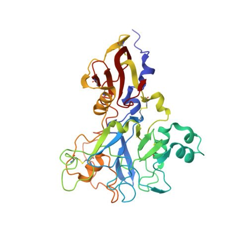
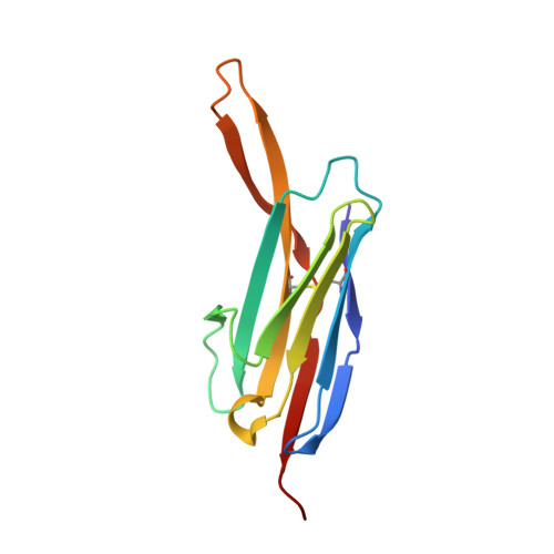
Structure of an IgNAR-AMA1 complex


Experimental Data Snapshot

  • Method: X-RAY DIFFRACTION
  • Resolution: 2.45 Å
  • R-Value Free: 
    0.282 (Depositor), 0.277 (DCC) 
  • R-Value Work: 
    0.189 (Depositor), 0.188 (DCC) 
  • R-Value Observed: 
    0.198 (Depositor) 

Starting Models: experimental
View more details

wwPDB Validation 3D Report Full Report

Validation slider image for 2Z8W

This is version 1.4 of the entry. See complete history. 

Literature

Structure of an IgNAR-AMA1 Complex: Targeting a Conserved Hydrophobic Cleft Broadens Malarial Strain Recognition

Henderson, K.A., Streltsov, V.A., Coley, A.M., Dolezal, O., Hudson, P.J., Batchelor, A.H., Gupta, A., Bai, T., Murphy, V.J., Anders, R.F., Foley, M., Nuttall, S.D.

(2007) Structure 15: 1452-1466

  • DOI: https://doi.org/10.1016/j.str.2007.09.011
  • Primary Citation Related Structures: 
    2Z8V, 2Z8W

  • PubMed Abstract: 

    Apical membrane antigen 1 (AMA1) is essential for invasion of erythrocytes and hepatocytes by Plasmodium parasites and is a leading malarial vaccine candidate. Although conventional antibodies to AMA1 can prevent such invasion, extensive polymorphisms within surface-exposed loops may limit the ability of these AMA1-induced antibodies to protect against all parasite genotypes. Using an AMA1-specific IgNAR single-variable-domain antibody, we performed targeted mutagenesis and selection against AMA1 from three P. falciparum strains. We present cocrystal structures of two antibody-AMA1 complexes which reveal extended IgNAR CDR3 loops penetrating deep into a hydrophobic cleft on the antigen surface and contacting residues conserved across parasite species. Comparison of a series of affinity-enhancing mutations allowed dissection of their relative contributions to binding kinetics and correlation with inhibition of erythrocyte invasion. These findings provide insights into mechanisms of single-domain antibody binding, and may enable design of reagents targeting otherwise cryptic epitopes in pathogen antigens.


  • Organizational Affiliation: 
    • CSIRO Molecular and Health Technologies, 343 Royal Parade, Parkville 3052, Australia.

Macromolecule Content 

  • Total Structure Weight: 102.44 kDa 
  • Atom Count: 7,674 
  • Modeled Residue Count: 902 
  • Deposited Residue Count: 902 
  • Unique protein chains: 2

Macromolecules

Find similar proteins by:|  3D Structure
Entity ID: 1
MoleculeChains  Sequence LengthOrganismDetailsImage
Apical membrane antigen 1
A, B
335Plasmodium falciparum 3D7Mutation(s): 0 
UniProt
Find proteins for Q7KQK5 (Plasmodium falciparum (isolate 3D7))
Explore Q7KQK5 
Go to UniProtKB:  Q7KQK5
Entity Groups
Sequence Clusters30% Identity50% Identity70% Identity90% Identity95% Identity100% Identity
UniProt GroupQ7KQK5
Sequence Annotations
Expand
Reference Sequence
Find similar proteins by:|  3D Structure
Entity ID: 2
MoleculeChains  Sequence LengthOrganismDetailsImage
New antigen receptor variable domain
C, D
116Orectolobus maculatusMutation(s): 2 
UniProt
Find proteins for Q6X1E6 (Orectolobus maculatus)
Explore Q6X1E6 
Go to UniProtKB:  Q6X1E6
Entity Groups
Sequence Clusters30% Identity50% Identity70% Identity90% Identity95% Identity100% Identity
UniProt GroupQ6X1E6
Sequence Annotations
Expand
Reference Sequence

Experimental Data & Validation

Experimental Data

  • Method: X-RAY DIFFRACTION
  • Resolution: 2.45 Å
  • R-Value Free:  0.282 (Depositor), 0.277 (DCC) 
  • R-Value Work:  0.189 (Depositor), 0.188 (DCC) 
  • R-Value Observed: 0.198 (Depositor) 
Space Group: P 31
Unit Cell:
Length ( Å )Angle ( ˚ )
a = 76.48α = 90
b = 76.48β = 90
c = 140.978γ = 120
Software Package:
Software NamePurpose
REFMACrefinement
ADSCdata collection
HKL-2000data reduction
HKL-2000data scaling
PHASERphasing

Structure Validation

View Full Validation Report


Entry History 

Deposition Data

Revision History  (Full details and data files)

  • Version 1.0: 2007-11-27
    Type: Initial release
  • Version 1.1: 2011-07-13
    Changes: Advisory, Source and taxonomy, Version format compliance
  • Version 1.2: 2021-11-10
    Changes: Database references
  • Version 1.3: 2023-11-01
    Changes: Data collection, Refinement description
  • Version 1.4: 2024-11-06
    Changes: Structure summary