2YXG | pdb_00002yxg

Crystal structure of Dihyrodipicolinate Synthase (dapA)


Experimental Data Snapshot

  • Method: X-RAY DIFFRACTION
  • Resolution: 2.20 Å
  • R-Value Free: 
    0.224 (Depositor) 
  • R-Value Work: 
    0.158 (Depositor) 
  • R-Value Observed: 
    0.161 (Depositor) 

Starting Model: experimental
View more details

wwPDB Validation 3D Report Full Report

Validation slider image for 2YXG

This is version 1.2 of the entry. See complete history

Literature

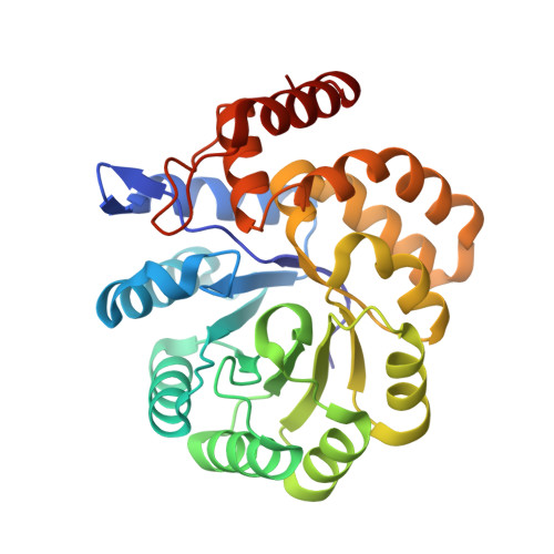
Structure of dihydrodipicolinate synthase from Methanocaldococcus jannaschii.

Padmanabhan, B.Strange, R.W.Antonyuk, S.V.Ellis, M.J.Hasnain, S.S.Iino, H.Agari, Y.Bessho, Y.Yokoyama, S.

(2009) Acta Crystallogr Sect F Struct Biol Cryst Commun 65: 1222-1226

  • DOI: https://doi.org/10.1107/S174430910904651X
  • Primary Citation Related Structures: 
    2YXG

  • PubMed Abstract: 

    In bacteria and plants, dihydrodipicolinate synthase (DHDPS) plays a key role in the (S)-lysine biosynthesis pathway. DHDPS catalyzes the first step of the condensation of (S)-aspartate-beta-semialdehyde and pyruvate to form an unstable compound, (4S)-4-hydroxy-2,3,4,5-tetrahydro-(2S)-dipicolinic acid. The activity of DHDPS is allosterically regulated by (S)-lysine, a feedback inhibitor. The crystal structure of DHDPS from Methanocaldococcus jannaschii (MjDHDPS) was solved by the molecular-replacement method and was refined to 2.2 A resolution. The structure revealed that MjDHDPS forms a functional homotetramer, as also observed in Escherichia coli DHDPS, Thermotoga maritima DHDPS and Bacillus anthracis DHDPS. The binding-site region of MjDHDPS is essentially similar to those found in other known DHDPS structures.


  • Organizational Affiliation
    • Systems and Structural Biology Center, Yokohama Institute, RIKEN, 1-7-22 Suehiro, Tsurumi, Yokohama 230-0045, Japan. bpadmanabhan@hotmail.com

Macromolecule Content 

  • Total Structure Weight: 126.45 kDa 
  • Atom Count: 9,549 
  • Modeled Residue Count: 1,153 
  • Deposited Residue Count: 1,156 
  • Unique protein chains: 1

Macromolecules

Find similar proteins by:|  3D Structure
Entity ID: 1
MoleculeChains  Sequence LengthOrganismDetailsImage
Dihydrodipicolinate synthase
A, B, C, D
289Methanocaldococcus jannaschii DSM 2661Mutation(s): 0 
Gene Names: dapA
EC: 4.2.1.52 (PDB Primary Data), 4.3.3.7 (UniProt)
UniProt
Find proteins for Q57695 (Methanocaldococcus jannaschii (strain ATCC 43067 / DSM 2661 / JAL-1 / JCM 10045 / NBRC 100440))
Explore Q57695 
Go to UniProtKB:  Q57695
Entity Groups
Sequence Clusters30% Identity50% Identity70% Identity90% Identity95% Identity100% Identity
UniProt GroupQ57695
Sequence Annotations
Expand
Reference Sequence

Experimental Data & Validation

Experimental Data

  • Method: X-RAY DIFFRACTION
  • Resolution: 2.20 Å
  • R-Value Free:  0.224 (Depositor) 
  • R-Value Work:  0.158 (Depositor) 
  • R-Value Observed: 0.161 (Depositor) 
Space Group: P 1 21 1
Unit Cell:
Length ( Å )Angle ( ˚ )
a = 80.469α = 90
b = 76.532β = 106.87
c = 101.863γ = 90
Software Package:
Software NamePurpose
REFMACrefinement
MAR345dtbdata collection
HKL-2000data reduction
SCALEPACKdata scaling
MOLREPphasing

Structure Validation

View Full Validation Report


Entry History 

Deposition Data

Revision History  (Full details and data files)

  • Version 1.0: 2007-10-30
    Type: Initial release
  • Version 1.1: 2011-07-13
    Changes: Version format compliance
  • Version 1.2: 2023-10-25
    Changes: Data collection, Database references, Refinement description