2YRV | pdb_00002yrv

Solution structure of the RBB1NT domain of human RB(retinoblastoma)-binding protein 1


Experimental Data Snapshot

  • Method: SOLUTION NMR
  • Conformers Calculated: 100 
  • Conformers Submitted: 20 
  • Selection Criteria: structures with the least restraint violations, target function 

wwPDB Validation 3D Report Full Report

Validation slider image for 2YRV

This is version 1.3 of the entry. See complete history

Literature

Solution structure of the RBB1NT domain of human RB(retinoblastoma)-binding protein 1

Nameki, N.Saito, K.Koshiba, S.Kigawa, T.Yokoyama, S.

To be published.

Macromolecule Content 

  • Total Structure Weight: 12.79 kDa 
  • Atom Count: 896 
  • Modeled Residue Count: 117 
  • Deposited Residue Count: 117 
  • Unique protein chains: 1

Macromolecules

Find similar proteins by:|  3D Structure
Entity ID: 1
MoleculeChains  Sequence LengthOrganismDetailsImage
AT-rich interactive domain-containing protein 4A117Homo sapiensMutation(s): 0 
Gene Names: ARID4A
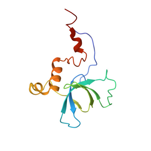
UniProt & NIH Common Fund Data Resources
Find proteins for P29374 (Homo sapiens)
Explore P29374 
Go to UniProtKB:  P29374
PHAROS:  P29374
GTEx:  ENSG00000032219 
Entity Groups
Sequence Clusters30% Identity50% Identity70% Identity90% Identity95% Identity100% Identity
UniProt GroupP29374
Sequence Annotations
Expand
Reference Sequence

Experimental Data & Validation

Experimental Data

  • Method: SOLUTION NMR
  • Conformers Calculated: 100 
  • Conformers Submitted: 20 
  • Selection Criteria: structures with the least restraint violations, target function 

Structure Validation

View Full Validation Report


Entry History 

Deposition Data

Revision History  (Full details and data files)

  • Version 1.0: 2008-04-08
    Type: Initial release
  • Version 1.1: 2011-07-13
    Changes: Version format compliance
  • Version 1.2: 2022-03-16
    Changes: Data collection, Database references, Derived calculations
  • Version 1.3: 2024-05-29
    Changes: Data collection