2YPH

Catalytic domain of mouse 2',3'-cyclic nucleotide 3'- phosphodiesterase, with mutation H230S, crystallized with 2',3-(RP)- cyclic-AMPS


Experimental Data Snapshot

  • Method: X-RAY DIFFRACTION
  • Resolution: 2.10 Å
  • R-Value Free: 0.289 
  • R-Value Work: 0.230 
  • R-Value Observed: 0.233 

wwPDB Validation   3D Report Full Report


Ligand Structure Quality Assessment 


This is version 2.2 of the entry. See complete history


Literature

Crystallographic Analysis of the Reaction Cycle of 2',3'-Cyclic Nucleotide 3'-Phosphodiesterase, a Unique Member of the 2H Phosphoesterase Family

Myllykoski, M.Raasakka, A.Lehtimaki, M.Han, H.Kursula, I.Kursula, P.

(2013) J Mol Biol 425: 4307

  • DOI: https://doi.org/10.1016/j.jmb.2013.06.012
  • Primary Citation of Related Structures:  
    2YOZ, 2YP0, 2YPC, 2YPE, 2YPH, 2YQ9, 3ZBR, 3ZBS, 3ZBZ

  • PubMed Abstract: 

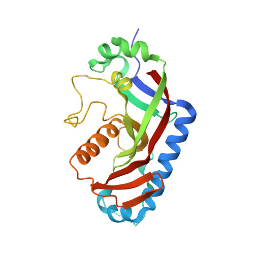
    2H phosphoesterases catalyze reactions on nucleotide substrates and contain two conserved histidine residues in the active site. Very limited information is currently available on the details of the active site and substrate/product binding during the catalytic cycle of these enzymes. We performed a comprehensive X-ray crystallographic study of mouse 2',3'-cyclic nucleotide 3'-phosphodiesterase (CNPase), a membrane-associated enzyme present at high levels in the tetrapod myelin sheath. We determined crystal structures of the CNPase phosphodiesterase domain complexed with substrate, product, and phosphorothioate analogues. The data provide detailed information on the CNPase reaction mechanism, including substrate binding mode and coordination of the nucleophilic water molecule. Linked to the reaction, an open/close motion of the β5-α7 loop is observed. The role of the N terminus of helix α7--unique for CNPase in the 2H family--during the reaction indicates that 2H phosphoesterases differ in their respective reaction mechanisms despite the conserved catalytic residues. Furthermore, based on small-angle X-ray scattering, we present a model for the full-length enzyme, indicating that the two domains of CNPase form an elongated molecule. Finally, based on our structural data and a comprehensive bioinformatics study, we discuss the conservation of CNPase in various organisms.


  • Organizational Affiliation

    Department of Biochemistry, University of Oulu, FIN-90014 Oulu, Finland; Biocenter Oulu, University of Oulu, FIN-90014 Oulu, Finland.


Macromolecules
Find similar proteins by:  (by identity cutoff)  |  3D Structure
Entity ID: 1
MoleculeChains Sequence LengthOrganismDetailsImage
2', 3'-CYCLIC-NUCLEOTIDE 3'-PHOSPHODIESTERASE221Mus musculusMutation(s): 1 
EC: 3.1.4.37
UniProt & NIH Common Fund Data Resources
Find proteins for P16330 (Mus musculus)
Explore P16330 
Go to UniProtKB:  P16330
IMPC:  MGI:88437
Entity Groups  
Sequence Clusters30% Identity50% Identity70% Identity90% Identity95% Identity100% Identity
UniProt GroupP16330
Sequence Annotations
Expand
  • Reference Sequence
Small Molecules
Ligands 3 Unique
IDChains Name / Formula / InChI Key2D Diagram3D Interactions
QQY
Query on QQY

Download Ideal Coordinates CCD File 
D [auth A][(3aR,4R,6R,6aR)-4-(6-aminopurin-9-yl)-2-oxidanylidene-2-sulfanyl-3a,4,6,6a-tetrahydrofuro[3,4-d][1,3,2]dioxaphosphol-6-yl]methanol
C10 H12 N5 O5 P S
OMSUUENXGWGOLC-PUHOFUEYSA-N
ACT
Query on ACT

Download Ideal Coordinates CCD File 
B [auth A]ACETATE ION
C2 H3 O2
QTBSBXVTEAMEQO-UHFFFAOYSA-M
CL
Query on CL

Download Ideal Coordinates CCD File 
C [auth A]CHLORIDE ION
Cl
VEXZGXHMUGYJMC-UHFFFAOYSA-M
Experimental Data & Validation

Experimental Data

  • Method: X-RAY DIFFRACTION
  • Resolution: 2.10 Å
  • R-Value Free: 0.289 
  • R-Value Work: 0.230 
  • R-Value Observed: 0.233 
  • Space Group: P 21 21 21
Unit Cell:
Length ( Å )Angle ( ˚ )
a = 40.63α = 90
b = 47.53β = 90
c = 107.55γ = 90
Software Package:
Software NamePurpose
PHENIXrefinement
XDSdata reduction
XSCALEdata scaling
PHASERphasing

Structure Validation

View Full Validation Report


Ligand Structure Quality Assessment 


Entry History 

Deposition Data

Revision History  (Full details and data files)

  • Version 1.0: 2013-07-10
    Type: Initial release
  • Version 1.1: 2013-07-17
    Changes: Database references
  • Version 1.2: 2013-11-13
    Changes: Database references
  • Version 2.0: 2018-01-17
    Changes: Atomic model, Data collection
  • Version 2.1: 2019-05-15
    Changes: Data collection, Experimental preparation
  • Version 2.2: 2023-12-20
    Changes: Data collection, Database references, Derived calculations, Other, Refinement description