2YPD | pdb_00002ypd

Crystal structure of the Jumonji domain of human Jumonji domain containing 1C protein


Experimental Data Snapshot

  • Method: X-RAY DIFFRACTION
  • Resolution: 2.10 Å
  • R-Value Free: 
    0.240 (Depositor), 0.243 (DCC) 
  • R-Value Work: 
    0.215 (Depositor), 0.220 (DCC) 
  • R-Value Observed: 
    0.216 (Depositor) 

wwPDB Validation 3D Report Full Report

Validation slider image for 2YPD

This is version 1.3 of the entry. See complete history

Literature

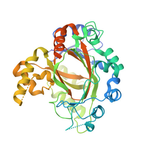
Crystal Structure of the Jumonji Domain of Human Jumonji Domain Containing 1C Protein

Vollmar, M.Johansson, C.Krojer, T.Strain-Damerell, C.Froese, S.Williams, E.Coutandin, D.von Delft, F.Weigelt, J.Arrowsmith, C.H.Bountra, C.Edwards, A.Oppermann, U.

To be published.

Macromolecule Content 

  • Total Structure Weight: 94.64 kDa 
  • Atom Count: 5,434 
  • Modeled Residue Count: 653 
  • Deposited Residue Count: 784 
  • Unique protein chains: 1

Macromolecules

Find similar proteins by:|  3D Structure
Entity ID: 1
MoleculeChains  Sequence LengthOrganismDetailsImage
PROBABLE JMJC DOMAIN-CONTAINING HISTONE DEMETHYLATION PROT EIN 2C
A, B
392Homo sapiensMutation(s): 0 
EC: 1.14.11
UniProt & NIH Common Fund Data Resources
Find proteins for Q15652 (Homo sapiens)
Explore Q15652 
Go to UniProtKB:  Q15652
PHAROS:  Q15652
GTEx:  ENSG00000171988 
Entity Groups
Sequence Clusters30% Identity50% Identity70% Identity90% Identity95% Identity100% Identity
UniProt GroupQ15652
Sequence Annotations
Expand
Reference Sequence

Small Molecules

Ligands 2 Unique
IDChains Name / Formula / InChI Key2D Diagram3D Interactions
EMT

Query on EMT


Download:Ideal Coordinates CCD File
C [auth A]
D [auth A]
E [auth A]
F [auth A]
G [auth A]
C [auth A],
D [auth A],
E [auth A],
F [auth A],
G [auth A],
H [auth A],
J [auth B],
K [auth B],
L [auth B],
M [auth B],
N [auth B]
2-(ETHYLMERCURI-THIO)-BENZOIC ACID
C9 H10 Hg O2 S
HXQVQGWHFRNKMS-UHFFFAOYSA-M
NA

Query on NA


Download:Ideal Coordinates CCD File
I [auth A],
O [auth B]
SODIUM ION
Na
FKNQFGJONOIPTF-UHFFFAOYSA-N

Experimental Data & Validation

Experimental Data

  • Method: X-RAY DIFFRACTION
  • Resolution: 2.10 Å
  • R-Value Free:  0.240 (Depositor), 0.243 (DCC) 
  • R-Value Work:  0.215 (Depositor), 0.220 (DCC) 
  • R-Value Observed: 0.216 (Depositor) 
Space Group: P 2 21 21
Unit Cell:
Length ( Å )Angle ( ˚ )
a = 45.718α = 90
b = 112.206β = 90
c = 165.173γ = 90
Software Package:
Software NamePurpose
REFMACrefinement
MOSFLMdata reduction
SCALAdata scaling
autoSHARPphasing

Structure Validation

View Full Validation Report


Entry History 

Revision History  (Full details and data files)

  • Version 1.0: 2012-12-12
    Type: Initial release
  • Version 1.1: 2014-09-17
    Changes: Atomic model, Derived calculations, Non-polymer description, Other, Source and taxonomy
  • Version 1.2: 2018-01-24
    Changes: Advisory, Database references
  • Version 1.3: 2024-05-08
    Changes: Data collection, Database references, Derived calculations, Other