2YNP

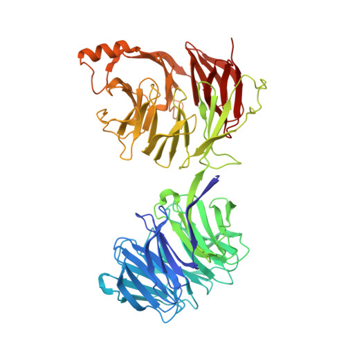
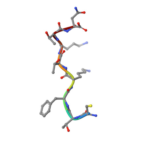
yeast betaprime COP 1-604 with KTKTN motif


Experimental Data Snapshot

  • Method: X-RAY DIFFRACTION
  • Resolution: 2.96 Å
  • R-Value Free: 0.294 
  • R-Value Work: 0.240 
  • R-Value Observed: 0.242 

Starting Models: experimental
View more details

wwPDB Validation   3D Report Full Report


This is version 1.3 of the entry. See complete history


Literature

Molecular Basis for Recognition of Dilysine Trafficking Motifs by Copi.

Jackson, L.P.Lewis, M.Kent, H.M.Edeling, M.A.Evans, P.R.Duden, R.Owen, D.J.

(2012) Dev Cell 23: 1255

  • DOI: https://doi.org/10.1016/j.devcel.2012.10.017
  • Primary Citation of Related Structures:  
    2YNN, 2YNO, 2YNP

  • PubMed Abstract: 

    COPI mediates retrograde trafficking from the Golgi to the endoplasmic reticulum (ER) and within the Golgi stack, sorting transmembrane proteins bearing C-terminal KKxx or KxKxx motifs. The structure of KxKxx motifs bound to the N-terminal WD-repeat domain of β'-COP identifies electrostatic contacts between the motif and complementary patches at the center of the β'-COP propeller. An absolute requirement of a two-residue spacing between the terminal carboxylate group and first lysine residue results from interactions of carbonyl groups in the motif backbone with basic side chains of β'-COP. Similar interactions are proposed to mediate binding of KKxx motifs by the homologous α-COP domain. Mutation of key interacting residues in either domain or in their cognate motifs abolishes in vitro binding and results in mistrafficking of dilysine-containing cargo in yeast without compromising cell viability. Flexibility between β'-COP WD-repeat domains and the location of cargo binding have implications for COPI coat assembly.


  • Organizational Affiliation

    Cambridge Institute for Medical Research, Department of Clinical Biochemistry, University of Cambridge, Hills Road, Cambridge CB2 0XY, UK. lpj21@cam.ac.uk


Macromolecules
Find similar proteins by:  (by identity cutoff)  |  3D Structure
Entity ID: 1
MoleculeChains Sequence LengthOrganismDetailsImage
COATOMER SUBUNIT BETA'604Saccharomyces cerevisiaeMutation(s): 0 
UniProt
Find proteins for P41811 (Saccharomyces cerevisiae (strain ATCC 204508 / S288c))
Explore P41811 
Go to UniProtKB:  P41811
Entity Groups  
Sequence Clusters30% Identity50% Identity70% Identity90% Identity95% Identity100% Identity
UniProt GroupP41811
Sequence Annotations
Expand
  • Reference Sequence

Find similar proteins by:  Sequence   |   3D Structure  

Entity ID: 2
MoleculeChains Sequence LengthOrganismDetailsImage
KTKTN MOTIFB [auth P]8synthetic constructMutation(s): 0 
Sequence Annotations
Expand
  • Reference Sequence
Experimental Data & Validation

Experimental Data

  • Method: X-RAY DIFFRACTION
  • Resolution: 2.96 Å
  • R-Value Free: 0.294 
  • R-Value Work: 0.240 
  • R-Value Observed: 0.242 
  • Space Group: P 31
Unit Cell:
Length ( Å )Angle ( ˚ )
a = 127.25α = 90
b = 127.25β = 90
c = 59.06γ = 120
Software Package:
Software NamePurpose
PHENIXrefinement
xia2data reduction
SCALAdata scaling
PHASERphasing

Structure Validation

View Full Validation Report


Entry History 

Deposition Data

Revision History  (Full details and data files)

  • Version 1.0: 2012-12-12
    Type: Initial release
  • Version 1.1: 2012-12-26
    Changes: Database references
  • Version 1.2: 2023-12-20
    Changes: Data collection, Database references, Other, Refinement description
  • Version 1.3: 2024-10-09
    Changes: Structure summary