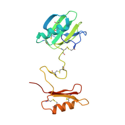
2YIL

Crystal Structure of Parasite Sarcocystis muris Lectin SML-2


Experimental Data Snapshot

  • Method: X-RAY DIFFRACTION
  • Resolution: 1.95 Å
  • R-Value Free: 0.197 
  • R-Value Work: 0.168 
  • R-Value Observed: 0.170 

wwPDB Validation   3D Report Full Report


This is version 1.5 of the entry. See complete history


Literature

Pan-Modular Structure of Microneme Protein Sml-2 from Parasite Sarcocystis Muris at 1.95 A Resolution and its Complex with 1-Thio-Beta-D-Galactose.

Mueller, J.J.Weiss, M.S.Heinemann, U.

(2011) Acta Crystallogr D Biol Crystallogr D67: 936

  • DOI: https://doi.org/10.1107/S0907444911037796
  • Primary Citation of Related Structures:  
    2YIL, 2YIO, 2YIP

  • PubMed Abstract: 

    The microneme protein SML-2 is a member of a small family of galactose-specific lectins that play a role during host-cell invasion by the apicomplexan parasite Sarcocystis muris. The structures of apo SML-2 and the 1-thio-β-D-galactose-SML-2 complex were determined at 1.95 and 2.1 Å resolution, respectively, by sulfur-SAD phasing. Highly elongated dimers are formed by PAN-domain tandems in the protomer, bearing the galactose-binding cavities at the distal apple-like domains. The detailed structure of the binding site in SML-2 explains the high specificity of galactose-endgroup binding and the broader specificity of the related Toxoplasma gondii protein TgMIC4 towards galactose and glucose. A large buried surface of highly hydrophobic character and 24 intersubunit hydrogen bonds stabilize the dimers and half of the 12 disulfides per dimer are shielded from the solvent by the polypeptide chain, thereby enhancing the resistance of the parasite protein towards unfolding and proteolysis that allows it to survive within the intestinal tracts of the intermediate and final hosts.


  • Organizational Affiliation

    Kristallographie, Max-Delbrück-Centrum für Molekulare Medizin, Robert-Rössle-Strasse 10, 13125 Berlin, Germany.


Macromolecules
Find similar proteins by:  (by identity cutoff)  |  3D Structure
Entity ID: 1
MoleculeChains Sequence LengthOrganismDetailsImage
MICRONEME ANTIGEN L2
A, B, C, D, E
A, B, C, D, E, F
138Sarcocystis murisMutation(s): 0 
UniProt
Find proteins for P81860 (Sarcocystis muris)
Explore P81860 
Go to UniProtKB:  P81860
Entity Groups  
Sequence Clusters30% Identity50% Identity70% Identity90% Identity95% Identity100% Identity
UniProt GroupP81860
Sequence Annotations
Expand
  • Reference Sequence
Small Molecules
Ligands 3 Unique
IDChains Name / Formula / InChI Key2D Diagram3D Interactions
SO4
Query on SO4

Download Ideal Coordinates CCD File 
M [auth C],
V [auth E],
W [auth E]
SULFATE ION
O4 S
QAOWNCQODCNURD-UHFFFAOYSA-L
GOL
Query on GOL

Download Ideal Coordinates CCD File 
H [auth A]
I [auth A]
K [auth B]
N [auth C]
O [auth C]
H [auth A],
I [auth A],
K [auth B],
N [auth C],
O [auth C],
P [auth C],
S [auth D],
T [auth D],
X [auth F],
Z [auth F]
GLYCEROL
C3 H8 O3
PEDCQBHIVMGVHV-UHFFFAOYSA-N
CL
Query on CL

Download Ideal Coordinates CCD File 
G [auth A]
J [auth B]
L [auth C]
Q [auth D]
R [auth D]
G [auth A],
J [auth B],
L [auth C],
Q [auth D],
R [auth D],
U [auth E],
Y [auth F]
CHLORIDE ION
Cl
VEXZGXHMUGYJMC-UHFFFAOYSA-M
Experimental Data & Validation

Experimental Data

  • Method: X-RAY DIFFRACTION
  • Resolution: 1.95 Å
  • R-Value Free: 0.197 
  • R-Value Work: 0.168 
  • R-Value Observed: 0.170 
  • Space Group: P 21 21 21
Unit Cell:
Length ( Å )Angle ( ˚ )
a = 53.17α = 90
b = 129.04β = 90
c = 158.1γ = 90
Software Package:
Software NamePurpose
REFMACrefinement
XDSdata reduction
XSCALEdata scaling
SHELXCDphasing
SHELXEphasing

Structure Validation

View Full Validation Report


Entry History 

Deposition Data

Revision History  (Full details and data files)

  • Version 1.0: 2011-11-02
    Type: Initial release
  • Version 1.1: 2011-11-30
    Changes: Database references
  • Version 1.2: 2019-01-30
    Changes: Data collection, Experimental preparation, Other
  • Version 1.3: 2019-02-06
    Changes: Data collection, Experimental preparation
  • Version 1.4: 2019-07-10
    Changes: Data collection
  • Version 1.5: 2019-07-17
    Changes: Data collection