2YHA | pdb_00002yha

Crystal Structure of the N. crassa QDE-2 AGO MID-PIWI Domains


Experimental Data Snapshot

  • Method: X-RAY DIFFRACTION
  • Resolution: 1.85 Å
  • R-Value Free: 
    0.236 (Depositor), 0.235 (DCC) 
  • R-Value Work: 
    0.196 (Depositor), 0.197 (DCC) 
  • R-Value Observed: 
    0.197 (Depositor) 

Starting Model: experimental
View more details

wwPDB Validation 3D Report Full Report

Validation slider image for 2YHA

This is version 1.3 of the entry. See complete history

Literature

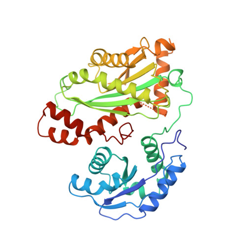
Crystal Structure of the Mid-Piwi Lobe of a Eukaryotic Argonaute Protein

Boland, A.Huntzinger, E.Schmidt, S.Izaurralde, E.Weichenriede, O.

(2011) Proc Natl Acad Sci U S A 108: 10466

  • DOI: https://doi.org/10.1073/pnas.1103946108
  • Primary Citation Related Structures: 
    2YHA, 2YHB

  • PubMed Abstract: 

    Argonaute proteins (AGOs) are essential effectors in RNA-mediated gene silencing pathways. They are characterized by a bilobal architecture, in which one lobe contains the N-terminal and PAZ domains and the other contains the MID and PIWI domains. Here, we present the first crystal structure of the MID-PIWI lobe from a eukaryotic AGO, the Neurospora crassa QDE-2 protein. Compared to prokaryotic AGOs, the domain orientation is conserved, indicating a conserved mode of nucleic acid binding. The PIWI domain shows an adaptable surface loop next to a eukaryote-specific α-helical insertion, which are both likely to contact the PAZ domain in a conformation-dependent manner to sense the functional state of the protein. The MID-PIWI interface is hydrophilic and buries residues that were previously thought to participate directly in the allosteric regulation of guide RNA binding. The interface includes the binding pocket for the guide RNA 5' end, and residues from both domains contribute to binding. Accordingly, micro-RNA (miRNA) binding is particularly sensitive to alteration in the MID-PIWI interface in Drosophila melanogaster AGO1 in vivo. The structure of the QDE-2 MID-PIWI lobe provides molecular and mechanistic insight into eukaryotic AGOs and has significant implications for understanding the role of these proteins in silencing.


  • Organizational Affiliation
    • Max Planck Institute for Developmental Biology, Spemannstrasse 35, D-72076 Tübingen, Germany.

Macromolecule Content 

  • Total Structure Weight: 43.17 kDa 
  • Atom Count: 3,065 
  • Modeled Residue Count: 368 
  • Deposited Residue Count: 388 
  • Unique protein chains: 1

Macromolecules

Find similar proteins by:|  3D Structure
Entity ID: 1
MoleculeChains  Sequence LengthOrganismDetailsImage
POST-TRANSCRIPTIONAL GENE SILENCING PROTEIN QDE-2388Neurospora crassaMutation(s): 0 
UniProt
Find proteins for Q9P8T1 (Neurospora crassa)
Explore Q9P8T1 
Go to UniProtKB:  Q9P8T1
Entity Groups
Sequence Clusters30% Identity50% Identity70% Identity90% Identity95% Identity100% Identity
UniProt GroupQ9P8T1
Sequence Annotations
Expand
Reference Sequence

Experimental Data & Validation

Experimental Data

  • Method: X-RAY DIFFRACTION
  • Resolution: 1.85 Å
  • R-Value Free:  0.236 (Depositor), 0.235 (DCC) 
  • R-Value Work:  0.196 (Depositor), 0.197 (DCC) 
  • R-Value Observed: 0.197 (Depositor) 
Space Group: P 32 2 1
Unit Cell:
Length ( Å )Angle ( ˚ )
a = 63.08α = 90
b = 63.08β = 90
c = 170.47γ = 120
Software Package:
Software NamePurpose
PHENIXrefinement
XDSdata reduction
XSCALEdata scaling
PHASERphasing

Structure Validation

View Full Validation Report


Entry History 

Deposition Data

Revision History  (Full details and data files)

  • Version 1.0: 2011-06-15
    Type: Initial release
  • Version 1.1: 2011-06-30
    Changes: Version format compliance
  • Version 1.2: 2011-07-13
    Changes: Version format compliance
  • Version 1.3: 2023-12-20
    Changes: Data collection, Database references, Derived calculations, Other, Refinement description