2YGY | pdb_00002ygy

Structure of wild type E. coli N-acetylneuraminic acid lyase in space group P21 crystal form II


Experimental Data Snapshot

  • Method: X-RAY DIFFRACTION
  • Resolution: 1.90 Å
  • R-Value Free: 
    0.224 (Depositor), 0.232 (DCC) 
  • R-Value Work: 
    0.194 (Depositor), 0.200 (DCC) 
  • R-Value Observed: 
    0.196 (Depositor) 

Starting Model: experimental
View more details

wwPDB Validation 3D Report Full Report

Validation slider image for 2YGY

This is version 1.2 of the entry. See complete history

Literature

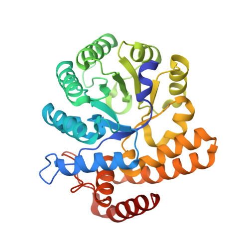
Pathological macromolecular crystallographic data affected by twinning, partial-disorder and exhibiting multiple lattices for testing of data processing and refinement tools.

Campeotto, I.Lebedev, A.Schreurs, A.M.M.Kroon-Batenburg, L.M.J.Lowe, E.Phillips, S.E.V.Murshudov, G.N.Pearson, A.R.

(2018) Sci Rep 8: 14876-14876

  • DOI: https://doi.org/10.1038/s41598-018-32962-6
  • Primary Citation Related Structures: 
    2YGY

  • PubMed Abstract: 

    Twinning is a crystal growth anomaly, which has posed a challenge in macromolecular crystallography (MX) since the earliest days. Many approaches have been used to treat twinned data in order to extract structural information. However, in most cases it is usually simpler to rescreen for new crystallization conditions that yield an untwinned crystal form or, if possible, collect data from non-twinned parts of the crystal. Here, we report 11 structures of engineered variants of the E. coli enzyme N-acetyl-neuraminic lyase which, despite twinning and incommensurate modulation, have been successfully indexed, solved and deposited. These structures span a resolution range of 1.45-2.30 Å, which is unusually high for datasets presenting such lattice disorders in MX and therefore these data provide an excellent test set for improving and challenging MX data processing programs.


  • Organizational Affiliation
    • Astbury Centre for Structural Molecular Biology, University of Leeds, Leeds, LS2 9JT, UK. ivan.campeotto@leicester.ac.uk.

Macromolecule Content 

  • Total Structure Weight: 134.96 kDa 
  • Atom Count: 9,923 
  • Modeled Residue Count: 1,189 
  • Deposited Residue Count: 1,216 
  • Unique protein chains: 1

Macromolecules

Find similar proteins by:|  3D Structure
Entity ID: 1
MoleculeChains  Sequence LengthOrganismDetailsImage
N-ACETYLNEURAMINATE LYASE
A, B, C, D
304Escherichia coli BL21(DE3)Mutation(s): 0 
EC: 4.1.3.3
UniProt
Find proteins for P0A6L4 (Escherichia coli (strain K12))
Explore P0A6L4 
Go to UniProtKB:  P0A6L4
Entity Groups
Sequence Clusters30% Identity50% Identity70% Identity90% Identity95% Identity100% Identity
UniProt GroupP0A6L4
Sequence Annotations
Expand
Reference Sequence

Experimental Data & Validation

Experimental Data

  • Method: X-RAY DIFFRACTION
  • Resolution: 1.90 Å
  • R-Value Free:  0.224 (Depositor), 0.232 (DCC) 
  • R-Value Work:  0.194 (Depositor), 0.200 (DCC) 
  • R-Value Observed: 0.196 (Depositor) 
Space Group: P 1 21 1
Unit Cell:
Length ( Å )Angle ( ˚ )
a = 84.26α = 90
b = 95.87β = 115.33
c = 91.38γ = 90
Software Package:
Software NamePurpose
REFMACrefinement
MOSFLMdata reduction
SCALAdata scaling
PHASERphasing

Structure Validation

View Full Validation Report


Entry History 

Deposition Data

Revision History  (Full details and data files)

  • Version 1.0: 2012-04-04
    Type: Initial release
  • Version 1.1: 2018-10-17
    Changes: Data collection, Database references
  • Version 1.2: 2023-12-20
    Changes: Data collection, Database references, Derived calculations, Other, Refinement description