2Y8C | pdb_00002y8c

Plasmodium falciparum dUTPase in complex with a trityl ligand


Experimental Data Snapshot

  • Method: X-RAY DIFFRACTION
  • Resolution: 2.10 Å
  • R-Value Free: 
    0.240 (Depositor), 0.240 (DCC) 
  • R-Value Work: 
    0.188 (Depositor), 0.187 (DCC) 
  • R-Value Observed: 
    0.191 (Depositor) 

Starting Model: experimental
View more details

wwPDB Validation 3D Report Full Report

Validation slider image for 2Y8C

Ligand Structure Quality Assessment 


This is version 1.1 of the entry. See complete history

Literature

Beta-Branched Acyclic Nucleoside Analogues as Inhibitors of Plasmodium Falciparum Dutpase

Baragana, B.Mccarthy, O.Sanchez, P.Bosch, C.Kaiser, M.Brun, R.Whittingham, J.L.Roberts, S.Zhou, X.-X.Wilson, K.S.Johansson, N.G.Gonzalez-Pacanowska, D.Gilbert, I.H.

(2011) Bioorg Med Chem 19: 2378

  • DOI: https://doi.org/10.1016/j.bmc.2011.02.012
  • Primary Citation Related Structures: 
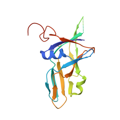
    2Y8C

  • PubMed Abstract: 

    We report a series of β-branched acyclic tritylated deoxyuridine analogues as inhibitors of Plasmodium falciparum deoxyuridine-5'-triphosphate nucleotidohydrolase (PfdUTPase), an enzyme involved in nucleotide metabolism that acts as first line of defence against uracil incorporation into DNA. Compounds were assayed against both PfdUTPase and intact parasites showing a correlation between enzyme inhibition and cellular assays. β-Branched acyclic uridine analogues described here showed equal or slightly better potency and selectivity compared with previously reported analogues. The best inhibitor gave a K(i) of 0.5 μM against PfdUTPase with selectivity greater than 200-fold compared to the corresponding human enzyme and sub-micromolar growth inhibition of P. falciparum (EC(50) 0.6 μM). A crystal structure of the complex of PfdUTPase with one of the inhibitors shows that this acyclic derivative binds to the active site in a similar manner to that previously reported for a tritylated cyclic deoxyuridine derivative.


  • Organizational Affiliation
    • Division of Biological Chemistry and Drug Discovery, College of Life Sciences, University of Dundee, Sir James Black Centre, Dundee DD1 5EH, UK.

Macromolecule Content 

  • Total Structure Weight: 60.54 kDa 
  • Atom Count: 3,399 
  • Modeled Residue Count: 404 
  • Deposited Residue Count: 519 
  • Unique protein chains: 1

Macromolecules

Find similar proteins by:|  3D Structure
Entity ID: 1
MoleculeChains  Sequence LengthOrganismDetailsImage
DEOXYURIDINE 5'-TRIPHOSPHATE NUCLEOTIDOHYDROLASE
A, B, C
173Plasmodium falciparum 3D7Mutation(s): 0 
EC: 3.6.1.23
UniProt
Find proteins for Q8II92 (Plasmodium falciparum (isolate 3D7))
Explore Q8II92 
Go to UniProtKB:  Q8II92
Entity Groups
Sequence Clusters30% Identity50% Identity70% Identity90% Identity95% Identity100% Identity
UniProt GroupQ8II92
Sequence Annotations
Expand
Reference Sequence

Experimental Data & Validation

Experimental Data

  • Method: X-RAY DIFFRACTION
  • Resolution: 2.10 Å
  • R-Value Free:  0.240 (Depositor), 0.240 (DCC) 
  • R-Value Work:  0.188 (Depositor), 0.187 (DCC) 
  • R-Value Observed: 0.191 (Depositor) 
Space Group: P 41
Unit Cell:
Length ( Å )Angle ( ˚ )
a = 77.16α = 90
b = 77.16β = 90
c = 110.98γ = 90
Software Package:
Software NamePurpose
REFMACrefinement
MOSFLMdata reduction
SCALAdata scaling
MOLREPphasing

Structure Validation

View Full Validation Report


Ligand Structure Quality Assessment 


Entry History 

Revision History  (Full details and data files)

  • Version 1.0: 2012-03-07
    Type: Initial release
  • Version 1.1: 2023-12-20
    Changes: Data collection, Database references, Derived calculations, Other, Refinement description