2Y83

Actin filament pointed end


Experimental Data Snapshot

  • Method: ELECTRON MICROSCOPY
  • Resolution: 22.9 Å
  • Aggregation State: FILAMENT 
  • Reconstruction Method: SINGLE PARTICLE 

wwPDB Validation   3D Report Full Report


This is version 1.2 of the entry. See complete history


Literature

Structural Basis for the Slow Dynamics of the Actin Filament Pointed End.

Narita, A.Oda, T.Maeda, Y.

(2011) EMBO J 30: 1230

  • DOI: https://doi.org/10.1038/emboj.2011.48
  • Primary Citation of Related Structures:  
    2Y83

  • PubMed Abstract: 

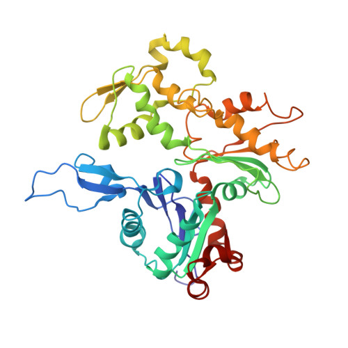
    The actin filament has clear polarity where one end, the pointed end, has a much slower polymerization and depolymerization rate than the other end, the barbed end. This intrinsic difference of the ends significantly affects all actin dynamics in the cell, which has central roles in a wide spectrum of cellular functions. The detailed mechanism underlying this difference has remained elusive, because high-resolution structures of the filament ends have not been available. Here, we present the structure of the actin filament pointed end obtained using a single particle analysis of cryo-electron micrographs. We determined that the terminal pointed end subunit is tilted towards the penultimate subunit, allowing specific and extra loop-to-loop inter-strand contacts between the two end subunits, which is not possible in other parts of the filament. These specific contacts prevent the end subunit from dissociating. For elongation, the loop-to-loop contacts also inhibit the incorporation of another actin monomer at the pointed end. These observations are likely to account for the less dynamic pointed end.


  • Organizational Affiliation

    Structural Biology Research Center and Division of Biological Science, Graduate School of Science, Nagoya University, Nagoya, Japan. narita.akihiro@f.mbox.nagoya-u.ac.jp


Macromolecules
Find similar proteins by:  (by identity cutoff)  |  3D Structure
Entity ID: 1
MoleculeChains Sequence LengthOrganismDetailsImage
ACTIN, ALPHA SKELETAL MUSCLE375Oryctolagus cuniculusMutation(s): 0 
UniProt
Find proteins for P68135 (Oryctolagus cuniculus)
Explore P68135 
Go to UniProtKB:  P68135
Entity Groups  
Sequence Clusters30% Identity50% Identity70% Identity90% Identity95% Identity100% Identity
UniProt GroupP68135
Sequence Annotations
Expand
  • Reference Sequence
Small Molecules
Ligands 2 Unique
IDChains Name / Formula / InChI Key2D Diagram3D Interactions
ADP
Query on ADP

Download Ideal Coordinates CCD File 
G [auth O]
I [auth P]
K [auth Q]
M [auth R]
O [auth S]
G [auth O],
I [auth P],
K [auth Q],
M [auth R],
O [auth S],
Q [auth T]
ADENOSINE-5'-DIPHOSPHATE
C10 H15 N5 O10 P2
XTWYTFMLZFPYCI-KQYNXXCUSA-N
CA
Query on CA

Download Ideal Coordinates CCD File 
H [auth O]
J [auth P]
L [auth Q]
N [auth R]
P [auth S]
H [auth O],
J [auth P],
L [auth Q],
N [auth R],
P [auth S],
R [auth T]
CALCIUM ION
Ca
BHPQYMZQTOCNFJ-UHFFFAOYSA-N
Experimental Data & Validation

Experimental Data

  • Method: ELECTRON MICROSCOPY
  • Resolution: 22.9 Å
  • Aggregation State: FILAMENT 
  • Reconstruction Method: SINGLE PARTICLE 
EM Software:
TaskSoftware PackageVersion
RECONSTRUCTIONEOS

Structure Validation

View Full Validation Report


Entry History 

Deposition Data

Revision History  (Full details and data files)

  • Version 1.0: 2011-03-30
    Type: Initial release
  • Version 1.1: 2013-03-20
    Changes: Other, Version format compliance
  • Version 1.2: 2018-10-03
    Changes: Data collection