2Y7L

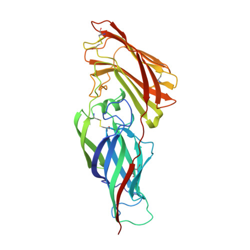
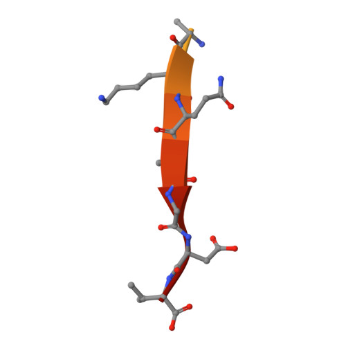
Structure of N-terminal domain of Candida albicans Als9-2 in complex with human fibrinogen gamma peptide


Experimental Data Snapshot

  • Method: X-RAY DIFFRACTION
  • Resolution: 1.49 Å
  • R-Value Free: 0.224 
  • R-Value Work: 0.196 
  • R-Value Observed: 0.198 

Starting Model: experimental
View more details

wwPDB Validation   3D Report Full Report


This is version 1.2 of the entry. See complete history


Literature

Structural Basis for the Broad Specificity to Host- Cell Ligands by the Pathogenic Fungus Candida Albicans.

Salgado, P.S.Yan, R.Taylor, J.D.Burchell, L.Jones, R.Hoyer, L.L.Matthews, S.J.Simpson, P.J.Cota, E.

(2011) Proc Natl Acad Sci U S A 108: 15775

  • DOI: https://doi.org/10.1073/pnas.1103496108
  • Primary Citation of Related Structures:  
    2Y7L, 2Y7M, 2Y7N, 2Y7O, 2YLH

  • PubMed Abstract: 

    Candida albicans is the most prevalent fungal pathogen in humans and a major source of life-threatening nosocomial infections. The Als (agglutinin-like sequence) glycoproteins are an important virulence factor for this fungus and have been associated with binding of host-cell surface proteins and small peptides of random sequence, the formation of biofilms and amyloid fibers. High-resolution structures of N-terminal Als adhesins (NT-Als; up to 314 amino acids) show that ligand recognition relies on a motif capable of binding flexible C termini of peptides in extended conformation. Central to this mechanism is an invariant lysine that recognizes the C-terminal carboxylate of ligands at the end of a deep-binding cavity. In addition to several protein-peptide interactions, a network of water molecules runs parallel to one side of the ligand and contributes to the recognition of diverse peptide sequences. These data establish NT-Als adhesins as a separate family of peptide-binding proteins and an unexpected adhesion system for primary, widespread protein-protein interactions at the Candida/host-cell interface.


  • Organizational Affiliation

    Division of Molecular Biosciences, Imperial College London, Exhibition Road, South Kensington SW7 2AZ, United Kingdom.


Macromolecules
Find similar proteins by:  (by identity cutoff)  |  3D Structure
Entity ID: 1
MoleculeChains Sequence LengthOrganismDetailsImage
AGGLUTININ-LIKE ALS9 PROTEIN312Candida albicansMutation(s): 0 
UniProt
Find proteins for A0A1D8PQ86 (Candida albicans (strain SC5314 / ATCC MYA-2876))
Explore A0A1D8PQ86 
Go to UniProtKB:  A0A1D8PQ86
Entity Groups  
Sequence Clusters30% Identity50% Identity70% Identity90% Identity95% Identity100% Identity
UniProt GroupA0A1D8PQ86
Sequence Annotations
Expand
  • Reference Sequence

Find similar proteins by:  Sequence   |   3D Structure  

Entity ID: 2
MoleculeChains Sequence LengthOrganismDetailsImage
FIBRINOGEN GAMMA CHAIN, ISOFORM CRA_A17Homo sapiensMutation(s): 0 
UniProt & NIH Common Fund Data Resources
Find proteins for P02679 (Homo sapiens)
Explore P02679 
Go to UniProtKB:  P02679
PHAROS:  P02679
GTEx:  ENSG00000171557 
Entity Groups  
Sequence Clusters30% Identity50% Identity70% Identity90% Identity95% Identity100% Identity
UniProt GroupP02679
Sequence Annotations
Expand
  • Reference Sequence
Experimental Data & Validation

Experimental Data

  • Method: X-RAY DIFFRACTION
  • Resolution: 1.49 Å
  • R-Value Free: 0.224 
  • R-Value Work: 0.196 
  • R-Value Observed: 0.198 
  • Space Group: P 21 21 21
Unit Cell:
Length ( Å )Angle ( ˚ )
a = 35.581α = 90
b = 69.273β = 90
c = 122.223γ = 90
Software Package:
Software NamePurpose
REFMACrefinement
DENZOdata reduction
SCALEPACKdata scaling
PHASERphasing

Structure Validation

View Full Validation Report


Entry History 

Deposition Data

Revision History  (Full details and data files)

  • Version 1.0: 2011-10-05
    Type: Initial release
  • Version 1.1: 2023-12-20
    Changes: Data collection, Database references, Other, Refinement description
  • Version 1.2: 2024-11-06
    Changes: Structure summary