2Y68 | pdb_00002y68

Structure-based design of a new series of D-glutamic acid-based inhibitors of bacterial MurD ligase


Experimental Data Snapshot

  • Method: X-RAY DIFFRACTION
  • Resolution: 1.49 Å
  • R-Value Free: 
    0.204 (Depositor), 0.206 (DCC) 
  • R-Value Work: 
    0.179 (Depositor), 0.181 (DCC) 
  • R-Value Observed: 
    0.181 (Depositor) 

Starting Model: experimental
View more details

wwPDB Validation 3D Report Full Report

Validation slider image for 2Y68

Ligand Structure Quality Assessment 


This is version 1.3 of the entry. See complete history

Literature

Structure-based design of a new series of D-glutamic acid based inhibitors of bacterial UDP-N-acetylmuramoyl-L-alanine:D-glutamate ligase (MurD).

Tomasic, T.Zidar, N.Sink, R.Kovac, A.Blanot, D.Contreras-Martel, C.Dessen, A.Muller-Premru, M.Zega, A.Gobec, S.Kikelj, D.Masic, L.P.

(2011) J Med Chem 54: 4600-4610

  • DOI: https://doi.org/10.1021/jm2002525
  • Primary Citation Related Structures: 
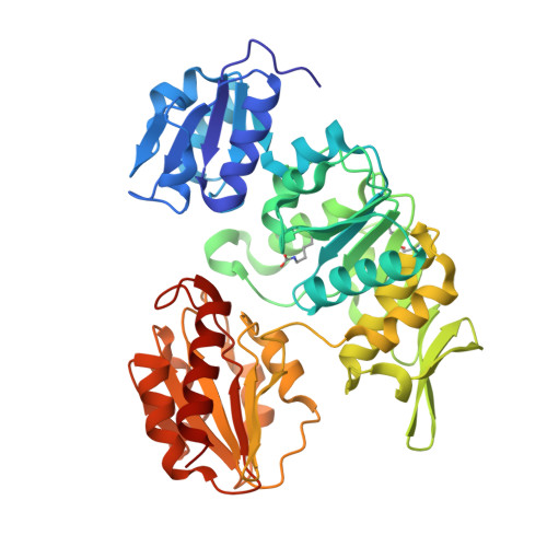
    2Y68

  • PubMed Abstract: 

    MurD ligase is one of the key enzymes participating in the intracellular steps of peptidoglycan biosynthesis and constitutes a viable target in the search for novel antibacterial drugs to combat bacterial drug-resistance. We have designed, synthesized, and evaluated a new series of D-glutamic acid-based Escherichia coli MurD inhibitors incorporating the 5-benzylidenethiazolidin-4-one scaffold. The crystal structure of 16 in the MurD active site has provided a good starting point for the design of structurally optimized inhibitors 73-75 endowed with improved MurD inhibitory potency (IC(50) between 3 and 7 μM). Inhibitors 74 and 75 showed weak activity against Gram-positive Staphylococcus aureus and Enterococcus faecalis. Compounds 73-75, with IC(50) values in the low micromolar range, represent the most potent D-Glu-based MurD inhibitors reported to date.


  • Organizational Affiliation
    • Faculty of Pharmacy, University of Ljubljana, Ljubljana, Slovenia.

Macromolecule Content 

  • Total Structure Weight: 49.31 kDa 
  • Atom Count: 3,900 
  • Modeled Residue Count: 435 
  • Deposited Residue Count: 445 
  • Unique protein chains: 1

Macromolecules

Find similar proteins by:|  3D Structure
Entity ID: 1
MoleculeChains  Sequence LengthOrganismDetailsImage
UDP-N-ACETYLMURAMOYLALANINE--D-GLUTAMATE LIGASE445Escherichia coli K-12Mutation(s): 0 
EC: 6.3.2.9
UniProt
Find proteins for P14900 (Escherichia coli (strain K12))
Explore P14900 
Go to UniProtKB:  P14900
Entity Groups
Sequence Clusters30% Identity50% Identity70% Identity90% Identity95% Identity100% Identity
UniProt GroupP14900
Sequence Annotations
Expand
Reference Sequence

Small Molecules

Ligands 5 Unique
IDChains Name / Formula / InChI Key2D Diagram3D Interactions
T04

Query on T04


Download:Ideal Coordinates CCD File
B [auth A]2-[[2-fluoro-5-[[[4-[(Z)-(4-oxo-2-sulfanylidene-1,3-thiazolidin-5-ylidene)methyl]phenyl]amino]methyl]phenyl]carbonylamino]pentanedioic acid
C23 H20 F N3 O6 S2
NEGUDZMAJDITEM-RMFUMNGISA-N
SO4

Query on SO4


Download:Ideal Coordinates CCD File
I [auth A],
J [auth A],
K [auth A],
L [auth A]
SULFATE ION
O4 S
QAOWNCQODCNURD-UHFFFAOYSA-L
DMS

Query on DMS


Download:Ideal Coordinates CCD File
C [auth A],
D [auth A]
DIMETHYL SULFOXIDE
C2 H6 O S
IAZDPXIOMUYVGZ-UHFFFAOYSA-N
AZI

Query on AZI


Download:Ideal Coordinates CCD File
E [auth A],
F [auth A],
G [auth A],
H [auth A]
AZIDE ION
N3
IVRMZWNICZWHMI-UHFFFAOYSA-N
CL

Query on CL


Download:Ideal Coordinates CCD File
M [auth A],
N [auth A],
O [auth A]
CHLORIDE ION
Cl
VEXZGXHMUGYJMC-UHFFFAOYSA-M
Modified Residues  1 Unique
IDChains TypeFormula2D DiagramParent
KCX
Query on KCX
A
L-PEPTIDE LINKINGC7 H14 N2 O4LYS

Experimental Data & Validation

Experimental Data

  • Method: X-RAY DIFFRACTION
  • Resolution: 1.49 Å
  • R-Value Free:  0.204 (Depositor), 0.206 (DCC) 
  • R-Value Work:  0.179 (Depositor), 0.181 (DCC) 
  • R-Value Observed: 0.181 (Depositor) 
Space Group: P 41
Unit Cell:
Length ( Å )Angle ( ˚ )
a = 65.544α = 90
b = 65.544β = 90
c = 134.959γ = 90
Software Package:
Software NamePurpose
REFMACrefinement
XDSdata reduction
XSCALEdata scaling
PHASERphasing

Structure Validation

View Full Validation Report


Ligand Structure Quality Assessment 


Entry History 

Revision History  (Full details and data files)

  • Version 1.0: 2011-06-08
    Type: Initial release
  • Version 1.1: 2011-08-31
    Changes: Database references, Version format compliance
  • Version 1.2: 2018-02-28
    Changes: Database references, Source and taxonomy
  • Version 1.3: 2023-12-20
    Changes: Data collection, Database references, Derived calculations, Other, Refinement description