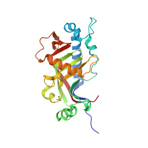
2Y1P

Catalytic domain of mouse 2',3'-cyclic nucleotide 3'- phosphodiesterase, complexed with citrate


Experimental Data Snapshot

  • Method: X-RAY DIFFRACTION
  • Resolution: 1.82 Å
  • R-Value Free: 0.199 
  • R-Value Work: 0.158 
  • R-Value Observed: 0.160 

Starting Model: experimental
View more details

wwPDB Validation   3D Report Full Report


This is version 2.0 of the entry. See complete history


Literature

Myelin 2',3'-Cyclic Nucleotide 3'-Phosphodiesterase: Active-Site Ligand Binding and Molecular Conformation.

Myllykoski, M.Raasakka, A.Han, H.Kursula, P.

(2012) PLoS One 7: 32336

  • DOI: https://doi.org/10.1371/journal.pone.0032336
  • Primary Citation of Related Structures:  
    2XMI, 2Y1P, 2Y3X, 2YDB, 2YDD

  • PubMed Abstract: 

    The 2',3'-cyclic nucleotide 3'-phosphodiesterase (CNPase) is a highly abundant membrane-associated enzyme in the myelin sheath of the vertebrate nervous system. CNPase is a member of the 2H phosphoesterase family and catalyzes the formation of 2'-nucleotide products from 2',3'-cyclic substrates; however, its physiological substrate and function remain unknown. It is likely that CNPase participates in RNA metabolism in the myelinating cell. We solved crystal structures of the phosphodiesterase domain of mouse CNPase, showing the binding mode of nucleotide ligands in the active site. The binding mode of the product 2'-AMP provides a detailed view of the reaction mechanism. Comparisons of CNPase crystal structures highlight flexible loops, which could play roles in substrate recognition; large differences in the active-site vicinity are observed when comparing more distant members of the 2H family. We also studied the full-length CNPase, showing its N-terminal domain is involved in RNA binding and dimerization. Our results provide a detailed picture of the CNPase active site during its catalytic cycle, and suggest a specific function for the previously uncharacterized N-terminal domain.


  • Organizational Affiliation

    Department of Biochemistry and Biocenter Oulu, University of Oulu, Oulu, Finland.


Macromolecules
Find similar proteins by:  (by identity cutoff)  |  3D Structure
Entity ID: 1
MoleculeChains Sequence LengthOrganismDetailsImage
2', 3'-CYCLIC NUCLEOTIDE 3'-PHOSPHODIESTERASE221Mus musculusMutation(s): 0 
EC: 3.1.4.37
UniProt & NIH Common Fund Data Resources
Find proteins for P16330 (Mus musculus)
Go to UniProtKB:  P16330
IMPC:  MGI:88437
Sequence Annotations
Expand
  • Reference Sequence
Small Molecules
Ligands 2 Unique
IDChains Name / Formula / InChI Key2D Diagram3D Interactions
CIT
Query on CIT

Download Ideal Coordinates CCD File 
B [auth A]CITRIC ACID
C6 H8 O7
KRKNYBCHXYNGOX-UHFFFAOYSA-N
GOL
Query on GOL

Download Ideal Coordinates CCD File 
C [auth A]GLYCEROL
C3 H8 O3
PEDCQBHIVMGVHV-UHFFFAOYSA-N
Experimental Data & Validation

Experimental Data

  • Method: X-RAY DIFFRACTION
  • Resolution: 1.82 Å
  • R-Value Free: 0.199 
  • R-Value Work: 0.158 
  • R-Value Observed: 0.160 
  • Space Group: P 1 21 1
Unit Cell:
Length ( Å )Angle ( ˚ )
a = 41.95α = 90
b = 46.92β = 95.51
c = 54.85γ = 90
Software Package:
Software NamePurpose
PHENIXrefinement
XDSdata reduction
XSCALEdata scaling
MOLREPphasing

Structure Validation

View Full Validation Report


Entry History 

Deposition Data

Revision History  (Full details and data files)

  • Version 1.0: 2011-12-21
    Type: Initial release
  • Version 1.1: 2012-03-21
    Changes: Other
  • Version 2.0: 2023-12-20
    Changes: Atomic model, Data collection, Database references, Derived calculations, Other, Refinement description