2Y1I

Crystal structure of a S-diastereomer analogue of the spore photoproduct in complex with fragment DNA polymerase I from Bacillus stearothermophilus


Experimental Data Snapshot

  • Method: X-RAY DIFFRACTION
  • Resolution: 2.78 Å
  • R-Value Free: 0.273 
  • R-Value Work: 0.226 
  • R-Value Observed: 0.228 

Starting Model: experimental
View more details

wwPDB Validation   3D Report Full Report


This is version 1.2 of the entry. See complete history


Literature

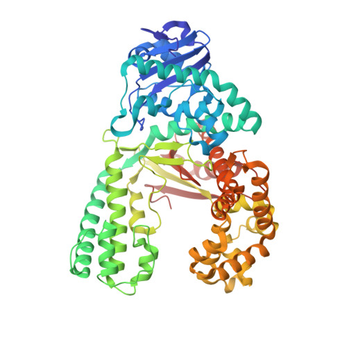
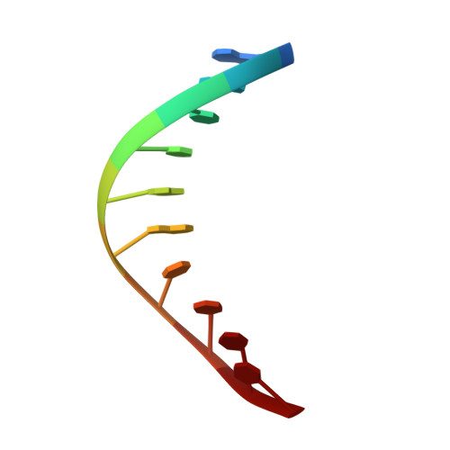
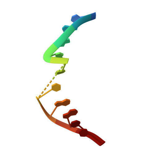
Crystal Structures and Repair Studies Reveal the Identity and the Base-Pairing Properties of the Uv-Induced Spore Photoproduct DNA Lesion.

Heil, K.Kneuttinger, A.C.Schneider, S.Lischke, U.Carell, T.

(2011) Chemistry 17: 9651

  • DOI: https://doi.org/10.1002/chem.201100177
  • Primary Citation of Related Structures:  
    2Y1I, 2Y1J

  • PubMed Abstract: 

    UV light is one of the major causes of DNA damage. In spore DNA, due to an unusual packing of the genetic material, a special spore photoproduct lesion (SP lesion) is formed, which is repaired by the enzyme spore photoproduct lyase (Spl), a radical S-adenosylmethionine (SAM) enzyme. We report here the synthesis and DNA incorporation of a DNA SP lesion analogue lacking the phosphodiester backbone. The oligonucleotides were used for repair studies and they were cocrystallized with a polymerase enzyme as a template to clarify the configuration of the SP lesion and to provide information about the base-pairing properties of the lesion. The structural analysis together with repair studies allowed us to clarify the identity of the preferentially repaired lesion diastereoisomer.


  • Organizational Affiliation

    Center for Integrated Protein Science (CiPSM) at the Department of Chemistry, Ludwig-Maximilians-University, Munich, Germany.


Macromolecules

Find similar proteins by:  (by identity cutoff)  |  3D Structure
Entity ID: 1
MoleculeChains Sequence LengthOrganismDetailsImage
DNA POLYMERASE I580Geobacillus stearothermophilusMutation(s): 0 
EC: 2.7.7.7
UniProt
Find proteins for E1C9K5 (Geobacillus stearothermophilus)
Explore E1C9K5 
Go to UniProtKB:  E1C9K5
Entity Groups  
Sequence Clusters30% Identity50% Identity70% Identity90% Identity95% Identity100% Identity
UniProt GroupE1C9K5
Sequence Annotations
Expand
  • Reference Sequence

Find similar nucleic acids by:  Sequence   |   3D Structure  

Entity ID: 2
MoleculeChains LengthOrganismImage
5'-D(*GP*AP*CP*CP*AP*AP*CP*CP*CP*TP)-3'10synthetic construct
Sequence Annotations
Expand
  • Reference Sequence

Find similar nucleic acids by:  Sequence   |   3D Structure  

Entity ID: 3
MoleculeChains LengthOrganismImage
5'-D(*AP*GP*GP*GP*PBTP*THM*GP*GP*TP*CP)-3'10synthetic construct
Sequence Annotations
Expand
  • Reference Sequence
Experimental Data & Validation

Experimental Data

  • Method: X-RAY DIFFRACTION
  • Resolution: 2.78 Å
  • R-Value Free: 0.273 
  • R-Value Work: 0.226 
  • R-Value Observed: 0.228 
  • Space Group: P 21 21 21
Unit Cell:
Length ( Å )Angle ( ˚ )
a = 88.669α = 90
b = 93.793β = 90
c = 107.101γ = 90
Software Package:
Software NamePurpose
REFMACrefinement
XDSdata reduction
SCALAdata scaling
PHASERphasing

Structure Validation

View Full Validation Report


Entry History 

Deposition Data

Revision History  (Full details and data files)

  • Version 1.0: 2011-07-27
    Type: Initial release
  • Version 1.1: 2011-11-02
    Changes: Database references
  • Version 1.2: 2023-12-20
    Changes: Data collection, Database references, Derived calculations, Other, Refinement description