2XZO | pdb_00002xzo

Upf1 helicase - RNA complex


Experimental Data Snapshot

  • Method: X-RAY DIFFRACTION
  • Resolution: 2.40 Å
  • R-Value Free: 
    0.233 (Depositor), 0.226 (DCC) 
  • R-Value Work: 
    0.193 (Depositor), 0.187 (DCC) 
  • R-Value Observed: 
    0.195 (Depositor) 

Starting Model: experimental
View more details

wwPDB Validation 3D Report Full Report

Validation slider image for 2XZO

Ligand Structure Quality Assessment 


This is version 1.4 of the entry. See complete history

Literature

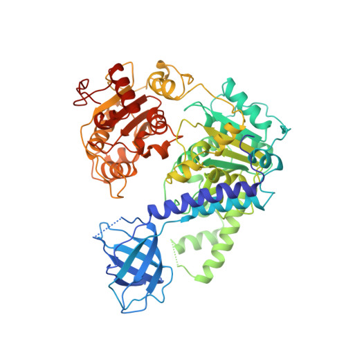
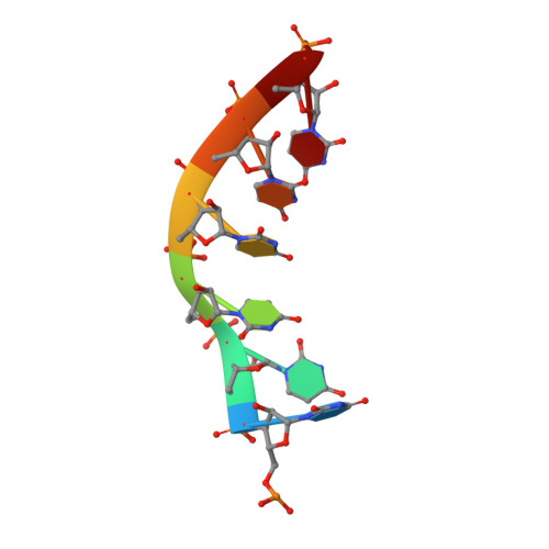
Molecular Mechanisms for the RNA-Dependent ATPase Activity of Upf1 and its Regulation by Upf2.

Chakrabarti, S.Jayachandran, U.Bonneau, F.Fiorini, F.Basquin, C.Domcke, S.Le Hir, H.Conti, E.

(2011) Mol Cell 41: 693

  • DOI: https://doi.org/10.1016/j.molcel.2011.02.010
  • Primary Citation Related Structures: 
    2XZL, 2XZO, 2XZP

  • PubMed Abstract: 

    Upf1 is a crucial factor in nonsense-mediated mRNA decay, the eukaryotic surveillance pathway that degrades mRNAs containing premature stop codons. The essential RNA-dependent ATPase activity of Upf1 is triggered by the formation of the surveillance complex with Upf2-Upf3. We report crystal structures of Upf1 in the presence and absence of the CH domain, captured in the transition state with ADP:AlF₄⁻ and RNA. In isolation, Upf1 clamps onto the RNA, enclosing it in a channel formed by both the catalytic and regulatory domains. Upon binding to Upf2, the regulatory CH domain of Upf1 undergoes a large conformational change, causing the catalytic helicase domain to bind RNA less extensively and triggering its helicase activity. Formation of the surveillance complex thus modifies the RNA binding properties and the catalytic activity of Upf1, causing it to switch from an RNA-clamping mode to an RNA-unwinding mode.


  • Organizational Affiliation
    • Max-Planck-Institute of Biochemistry, Department of Structural Cell Biology, Am Klopferspitz 18, D-82152 Martinsried, Germany.

Macromolecule Content 

  • Total Structure Weight: 73.42 kDa 
  • Atom Count: 4,932 
  • Modeled Residue Count: 620 
  • Deposited Residue Count: 630 
  • Unique protein chains: 1
  • Unique nucleic acid chains: 1

Macromolecules


Find similar proteins by:|  3D Structure
Entity ID: 1
MoleculeChains  Sequence LengthOrganismDetailsImage
REGULATOR OF NONSENSE TRANSCRIPTS 1623Homo sapiensMutation(s): 0 
EC: 3.6.4.13 (PDB Primary Data), 3.6.4.12 (UniProt)
UniProt & NIH Common Fund Data Resources
Find proteins for Q92900 (Homo sapiens)
Explore Q92900 
Go to UniProtKB:  Q92900
PHAROS:  Q92900
GTEx:  ENSG00000005007 
Entity Groups
Sequence Clusters30% Identity50% Identity70% Identity90% Identity95% Identity100% Identity
UniProt GroupQ92900
Sequence Annotations
Expand
Reference Sequence
Find similar nucleic acids by:  Sequence
Entity ID: 2
MoleculeChains LengthOrganismImage
5'-R(*UP*UP*UP*UP*UP*UP*UP)-3'B [auth D]7synthetic construct
Sequence Annotations
Expand
Reference Sequence

Small Molecules

Ligands 4 Unique
IDChains Name / Formula / InChI Key2D Diagram3D Interactions
ADP

Query on ADP


Download:Ideal Coordinates CCD File
F [auth A]ADENOSINE-5'-DIPHOSPHATE
C10 H15 N5 O10 P2
XTWYTFMLZFPYCI-KQYNXXCUSA-N
1PE

Query on 1PE


Download:Ideal Coordinates CCD File
C [auth A],
D [auth A],
E [auth A]
PENTAETHYLENE GLYCOL
C10 H22 O6
JLFNLZLINWHATN-UHFFFAOYSA-N
ALF

Query on ALF


Download:Ideal Coordinates CCD File
G [auth A]TETRAFLUOROALUMINATE ION
Al F4
UYOMQIYKOOHAMK-UHFFFAOYSA-J
MG

Query on MG


Download:Ideal Coordinates CCD File
H [auth A],
I [auth A]
MAGNESIUM ION
Mg
JLVVSXFLKOJNIY-UHFFFAOYSA-N

Experimental Data & Validation

Experimental Data

  • Method: X-RAY DIFFRACTION
  • Resolution: 2.40 Å
  • R-Value Free:  0.233 (Depositor), 0.226 (DCC) 
  • R-Value Work:  0.193 (Depositor), 0.187 (DCC) 
  • R-Value Observed: 0.195 (Depositor) 
Space Group: P 21 21 21
Unit Cell:
Length ( Å )Angle ( ˚ )
a = 77.066α = 90
b = 101.896β = 90
c = 112.32γ = 90
Software Package:
Software NamePurpose
PHENIXrefinement
XDSdata reduction
SCALAdata scaling
PHASERphasing

Structure Validation

View Full Validation Report


Ligand Structure Quality Assessment 


Entry History 

Deposition Data

Revision History  (Full details and data files)

  • Version 1.0: 2011-03-30
    Type: Initial release
  • Version 1.1: 2011-05-08
    Changes: Version format compliance
  • Version 1.2: 2011-07-13
    Changes: Version format compliance
  • Version 1.3: 2017-07-12
    Changes: Advisory, Derived calculations
  • Version 1.4: 2023-12-20
    Changes: Data collection, Database references, Other, Refinement description