2XYU | pdb_00002xyu

Crystal structure of EphA4 kinase domain in complex with VUF 12058


Experimental Data Snapshot

  • Method: X-RAY DIFFRACTION
  • Resolution: 2.12 Å
  • R-Value Free: 
    0.230 (Depositor) 
  • R-Value Work: 
    0.189 (Depositor), 0.199 (DCC) 
  • R-Value Observed: 
    0.192 (Depositor) 

Starting Model: experimental
View more details

wwPDB Validation 3D Report Full Report

Validation slider image for 2XYU

Ligand Structure Quality Assessment 


This is version 1.4 of the entry. See complete history

Literature

Fragment Based Lead Discovery of Small Molecule Inhibitors for the Epha4 Receptor Tyrosine Kinase.

Van Linden, O.P.Farenc, C.J.A.Zoutman, W.H.Hameetman, L.Wijtmans, M.Leurs, R.Tensen, C.P.Siegal, G.De Esch, I.J.

(2012) Eur J Med Chem 47: 493

  • DOI: https://doi.org/10.1016/j.ejmech.2011.11.020
  • Primary Citation Related Structures: 
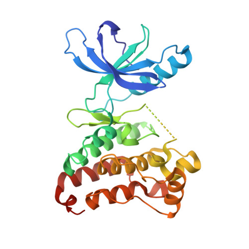
    2XYU

  • PubMed Abstract: 

    The in silico identification, optimization and crystallographic characterization of a 6,7,8,9-tetrahydro-3H-pyrazolo[3,4-c]isoquinolin-1-amine scaffold as an inhibitor for the EPHA4 receptor tyrosine kinase is described. A database containing commercially available compounds was subjected to an in silico screening procedure which was focused on finding novel, EPHA4 hinge binding fragments. This resulted in the identification of 6,7,8,9-tetrahydro-3H-pyrazolo[3,4-c]isoquinolin-1-amine derivatives as EPHA4 inhibitors. Hit exploration yielded a compound with 2 μM (IC(50)) affinity for the EPHA4 receptor tyrosine kinase domain. Soaking experiments into a crystal of the EPHA4 kinase domain gave a 2.11Å X-ray structure of the EPHA4 - inhibitor complex, which confirmed the binding mode of the scaffold as proposed by the initial in silico work. The results underscore the strength of fragment based in silico screening as a tool for the discovery of novel lead compounds as small molecule kinase inhibitors.


  • Organizational Affiliation
    • Leiden/Amsterdam Center for Drug Research (LACDR), Division of Medicinal Chemistry, Faculty of Sciences, VU University Amsterdam, De Boelelaan 1083, 1081 HV Amsterdam, The Netherlands.

Macromolecule Content 

  • Total Structure Weight: 33.08 kDa 
  • Atom Count: 2,224 
  • Modeled Residue Count: 260 
  • Deposited Residue Count: 285 
  • Unique protein chains: 1

Macromolecules

Find similar proteins by:|  3D Structure
Entity ID: 1
MoleculeChains  Sequence LengthOrganismDetailsImage
EPHRIN TYPE-A RECEPTOR 4,285Mus musculusMutation(s): 0 
EC: 2.7.10.1
UniProt
Find proteins for Q03137 (Mus musculus)
Explore Q03137 
Go to UniProtKB:  Q03137
Entity Groups
Sequence Clusters30% Identity50% Identity70% Identity90% Identity95% Identity100% Identity
UniProt GroupQ03137
Sequence Annotations
Expand
Reference Sequence

Small Molecules

Ligands 2 Unique
IDChains Name / Formula / InChI Key2D Diagram3D Interactions
Q9G

Query on Q9G


Download:Ideal Coordinates CCD File
H [auth A]5-(5-FLUORO-2-METHYLPHENYL)-6,7,8,9-TETRAHYDRO-3H-PYRAZOLO[3,4-C]ISOQUINOLIN-1-AMINE
C17 H17 F N4
YPULTKAHHAYKOE-UHFFFAOYSA-N
GOL

Query on GOL


Download:Ideal Coordinates CCD File
B [auth A]
C [auth A]
D [auth A]
E [auth A]
F [auth A]
B [auth A],
C [auth A],
D [auth A],
E [auth A],
F [auth A],
G [auth A]
GLYCEROL
C3 H8 O3
PEDCQBHIVMGVHV-UHFFFAOYSA-N
Binding Affinity Annotations 
IDSourceBinding Affinity
Q9G BindingDB:  2XYU IC50: 4500 (nM) from 1 assay(s)

Experimental Data & Validation

Experimental Data

  • Method: X-RAY DIFFRACTION
  • Resolution: 2.12 Å
  • R-Value Free:  0.230 (Depositor) 
  • R-Value Work:  0.189 (Depositor), 0.199 (DCC) 
  • R-Value Observed: 0.192 (Depositor) 
Space Group: P 21 21 21
Unit Cell:
Length ( Å )Angle ( ˚ )
a = 32.544α = 90
b = 91.545β = 90
c = 97.959γ = 90
Software Package:
Software NamePurpose
PHENIXrefinement
XDSdata reduction
XDSdata scaling
AMoREphasing

Structure Validation

View Full Validation Report


Ligand Structure Quality Assessment 


Entry History 

Deposition Data

Revision History  (Full details and data files)

  • Version 1.0: 2011-11-30
    Type: Initial release
  • Version 1.1: 2011-12-28
    Changes: Other
  • Version 1.2: 2012-01-25
    Changes: Other
  • Version 1.3: 2012-02-01
    Changes: Other
  • Version 1.4: 2023-12-20
    Changes: Data collection, Database references, Derived calculations, Other, Refinement description