2XRQ | pdb_00002xrq

Crystal structures exploring the origins of the broader specificity of escherichia coli heat-labile enterotoxin compared to cholera toxin


Experimental Data Snapshot

  • Method: X-RAY DIFFRACTION
  • Resolution: 2.40 Å
  • R-Value Free: 
    0.235 (Depositor), 0.236 (DCC) 
  • R-Value Work: 
    0.212 (Depositor) 
  • R-Value Observed: 
    0.213 (Depositor) 

Starting Model: experimental
View more details

wwPDB Validation 3D Report Full Report

Validation slider image for 2XRQ

This is version 2.2 of the entry. See complete history

Literature

Crystal Structures Exploring the Origins of the Broader Specificity of Escherichia Coli Heat-Labile Enterotoxin Compared to Cholera Toxin

Holmner, A.Mackenzie, A.Okvist, M.Jansson, L.Lebens, M.Teneberg, S.Krengel, U.

(2011) J Mol Biology 406: 387

  • DOI: https://doi.org/10.1016/j.jmb.2010.11.060
  • Primary Citation Related Structures: 
    2XRQ, 2XRS

  • PubMed Abstract: 

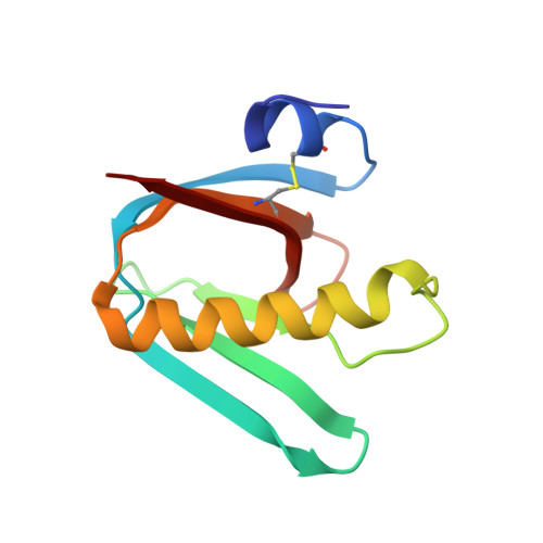
    Cholera toxin (CT) and Escherichia coli heat-labile enterotoxin (LT) are structurally and functionally related and share the same primary receptor, the GM1 ganglioside. Despite their extensive similarities, these two toxins exhibit distinct ligand specificities, with LT being more promiscuous than CT. Here, we have attempted to rationalize the broader binding specificity of LT and the subtle differences between the binding characteristics of LTs from human and porcine origins (mediated by their B subunit pentamers, hLTB and pLTB, respectively). The analysis is based on two crystal structures of pLTB in complexes with the pentasaccharide of its primary ligand, GM1, and with neolactotetraose, the carbohydrate determinant of a typical secondary ligand of LTs, respectively. Important molecular determinants underlying the different binding specificities of LTB and CTB are found to be contributed by Ser95, Tyr18 and Thr4 (or Ser4 of hLTB), which together prestabilize the binding site by positioning Lys91, Glu51 and the adjacent loop region (50-61) containing Ile58 for ligand binding. Glu7 and Ala1 may also play an important role. Many of these residues are closely connected with a recently identified second binding site, and there appears to be cross-talk between the two sites. Binding to N-acetyllactosamine-terminated receptors is further augmented by Arg13 (present in pLT and some hLT variants), as previously predicted.


  • Organizational Affiliation
    • Department of Chemistry, University of Oslo, P.O. Box 1033 Blindern, NO-0315 Oslo, Norway. asa.holmner-rocklov@vll.se

Macromolecule Content 

  • Total Structure Weight: 64.03 kDa 
  • Atom Count: 4,576 
  • Modeled Residue Count: 515 
  • Deposited Residue Count: 515 
  • Unique protein chains: 1

Macromolecules

Find similar proteins by:|  3D Structure
Entity ID: 1
MoleculeChains  Sequence LengthOrganismDetailsImage
HEAT-LABILE ENTEROTOXIN B CHAINA [auth D],
B [auth E],
C [auth F],
D [auth G],
E [auth H]
103Escherichia coliMutation(s): 0 
UniProt
Find proteins for P32890 (Escherichia coli)
Explore P32890 
Go to UniProtKB:  P32890
Entity Groups
Sequence Clusters30% Identity50% Identity70% Identity90% Identity95% Identity100% Identity
UniProt GroupP32890
Sequence Annotations
Expand
Reference Sequence

Oligosaccharides

Help  
Entity ID: 2
MoleculeChains Length2D Diagram GlycosylationD Interactions
beta-D-galactopyranose-(1-3)-2-acetamido-2-deoxy-beta-D-galactopyranose-(1-4)-[N-acetyl-alpha-neuraminic acid-(2-3)]beta-D-galactopyranose-(1-4)-beta-D-glucopyranoseF [auth A],
G [auth B],
H [auth C],
I,
J
5N/A
Glycosylation Resources
GlyTouCan: G48558GR
GlyCosmos: G48558GR
GlyGen: G48558GR

Experimental Data & Validation

Experimental Data

  • Method: X-RAY DIFFRACTION
  • Resolution: 2.40 Å
  • R-Value Free:  0.235 (Depositor), 0.236 (DCC) 
  • R-Value Work:  0.212 (Depositor) 
  • R-Value Observed: 0.213 (Depositor) 
Space Group: C 1 2 1
Unit Cell:
Length ( Å )Angle ( ˚ )
a = 112.009α = 90
b = 96.099β = 97.22
c = 66.289γ = 90
Software Package:
Software NamePurpose
REFMACrefinement
MOSFLMdata reduction
SCALAdata scaling
MOLREPphasing

Structure Validation

View Full Validation Report


Entry History 

Deposition Data

Revision History  (Full details and data files)

  • Version 1.0: 2010-12-08
    Type: Initial release
  • Version 1.1: 2012-06-06
    Changes: Database references, Version format compliance
  • Version 1.2: 2015-04-01
    Changes: Database references
  • Version 2.0: 2020-07-29
    Type: Remediation
    Reason: Carbohydrate remediation
    Changes: Advisory, Atomic model, Data collection, Derived calculations, Other, Structure summary
  • Version 2.1: 2023-12-20
    Changes: Data collection, Database references, Refinement description, Structure summary
  • Version 2.2: 2024-10-16
    Changes: Structure summary