


Myosin 1E SH3
Allsop, G., Harris, S.A., Peckham, M., Edwards, T.To be published.
Experimental Data Snapshot
wwPDB Validation 3D Report Full Report

Entity ID: 1 | |||||
|---|---|---|---|---|---|
| Molecule | Chains | Sequence Length | Organism | Details | Image |
| MYOSIN 1E SH3 | 60 | Mus musculus | Mutation(s): 0 | 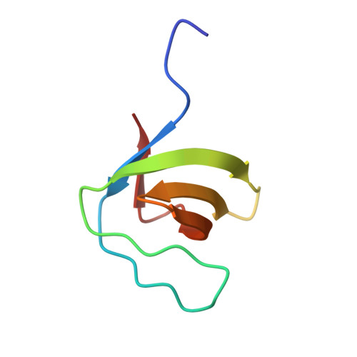 | |
UniProt | |||||
Entity Groups | |||||
| Sequence Clusters | 30% Identity50% Identity70% Identity90% Identity95% Identity100% Identity | ||||
| UniProt Group | E9Q634 | ||||
Sequence AnnotationsExpand | |||||
Reference Sequence | |||||
| Ligands 1 Unique | |||||
|---|---|---|---|---|---|
| ID | Chains | Name / Formula / InChI Key | 2D Diagram | 3D Interactions | |
| DIA Download:Ideal Coordinates CCD File | B [auth A] | OCTANE 1,8-DIAMINE C8 H20 N2 PWGJDPKCLMLPJW-UHFFFAOYSA-N |  | ||
| Length ( Å ) | Angle ( ˚ ) |
|---|---|
| a = 43.562 | α = 90 |
| b = 55.56 | β = 90 |
| c = 42.176 | γ = 90 |
| Software Name | Purpose |
|---|---|
| REFMAC | refinement |


