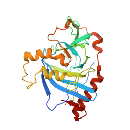
2XLA

Structure and metal-loading of a soluble periplasm cupro-protein: Cu- CucA-closed


Experimental Data Snapshot

  • Method: X-RAY DIFFRACTION
  • Resolution: 1.93 Å
  • R-Value Free: 0.203 
  • R-Value Work: 0.161 
  • R-Value Observed: 0.163 

Starting Model: experimental
View more details

wwPDB Validation   3D Report Full Report


This is version 1.4 of the entry. See complete history


Literature

Structure and Metal Loading of a Soluble Periplasm Cuproprotein.

Waldron, K.J.Firbank, S.J.Dainty, S.J.Perez-Rama, M.Tottey, S.Robinson, N.J.

(2010) J Biol Chem 285: 32504

  • DOI: https://doi.org/10.1074/jbc.M110.153080
  • Primary Citation of Related Structures:  
    2XL7, 2XL9, 2XLA, 2XLF, 2XLG

  • PubMed Abstract: 

    A copper-trafficking pathway was found to enable Cu(2+) occupancy of a soluble periplasm protein, CucA, even when competing Zn(2+) is abundant in the periplasm. Here, we solved the structure of CucA (a new cupin) and found that binding of Cu(2+), but not Zn(2+), quenches the fluorescence of Trp(165), which is adjacent to the metal site. Using this fluorescence probe, we established that CucA becomes partly occupied by Zn(2+) following exposure to equimolar Zn(2+) and Cu(2+). Cu(2+)-CucA is more thermodynamically stable than Zn(2+)-CucA but k((Zn→Cu)exchange) is slow, raising questions about how the periplasm contains solely the Cu(2+) form. We discovered that a copper-trafficking pathway involving two copper transporters (CtaA and PacS) and a metallochaperone (Atx1) is obligatory for Cu(2+)-CucA to accumulate in the periplasm. There was negligible CucA protein in the periplasm of ΔctaA cells, but the abundance of cucA transcripts was unaltered. Crucially, ΔctaA cells overaccumulate low M(r) copper complexes in the periplasm, and purified apoCucA can readily acquire Cu(2+) from ΔctaA periplasm extracts, but in vivo apoCucA fails to come into contact with these periplasmic copper pools. Instead, copper traffics via a cytoplasmic pathway that is coupled to CucA translocation to the periplasm.


  • Organizational Affiliation

    Institute for Cell and Molecular Biosciences, University of Newcastle Medical School, Newcastle upon Tyne NE2 4HH, United Kingdom.


Macromolecules
Find similar proteins by:  (by identity cutoff)  |  3D Structure
Entity ID: 1
MoleculeChains Sequence LengthOrganismDetailsImage
SLL1785 PROTEIN
A, B, C, D
239Synechocystis sp. PCC 6803Mutation(s): 0 
UniProt
Find proteins for P73600 (Synechocystis sp. (strain ATCC 27184 / PCC 6803 / Kazusa))
Explore P73600 
Go to UniProtKB:  P73600
Entity Groups  
Sequence Clusters30% Identity50% Identity70% Identity90% Identity95% Identity100% Identity
UniProt GroupP73600
Sequence Annotations
Expand
  • Reference Sequence
Experimental Data & Validation

Experimental Data

  • Method: X-RAY DIFFRACTION
  • Resolution: 1.93 Å
  • R-Value Free: 0.203 
  • R-Value Work: 0.161 
  • R-Value Observed: 0.163 
  • Space Group: P 21 21 21
Unit Cell:
Length ( Å )Angle ( ˚ )
a = 50.31α = 90
b = 72.51β = 90
c = 262.1γ = 90
Software Package:
Software NamePurpose
REFMACrefinement
MOSFLMdata reduction
SCALAdata scaling
MOLREPphasing

Structure Validation

View Full Validation Report


Entry History 

Deposition Data

Revision History  (Full details and data files)

  • Version 1.0: 2010-08-11
    Type: Initial release
  • Version 1.1: 2011-05-12
    Changes: Version format compliance
  • Version 1.2: 2011-07-13
    Changes: Version format compliance
  • Version 1.3: 2019-09-25
    Changes: Data collection, Experimental preparation, Other
  • Version 1.4: 2023-12-20
    Changes: Data collection, Database references, Derived calculations, Refinement description