2XKN | pdb_00002xkn

Crystal structure of the Fab fragment of the anti-EGFR antibody 7A7


Experimental Data Snapshot

  • Method: X-RAY DIFFRACTION
  • Resolution: 1.40 Å
  • R-Value Free: 
    0.176 (Depositor), 0.179 (DCC) 
  • R-Value Work: 
    0.144 (Depositor), 0.149 (DCC) 
  • R-Value Observed: 
    0.145 (Depositor) 

Starting Model: experimental
View more details

wwPDB Validation 3D Report Full Report

Validation slider image for 2XKN

This is version 1.3 of the entry. See complete history

Literature

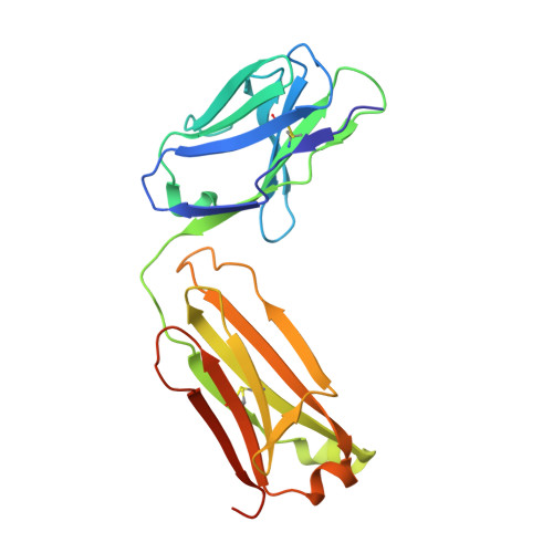
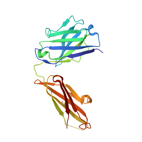
Structure of the Fab Fragment of the Anti-Murine Egfr Antibody 7A7 and Exploration of its Receptor Binding Site.

Talavera, A.Mackenzie, J.Garrido, G.Friemann, R.Lopez-Requena, A.Moreno, E.Krengel, U.

(2011) Mol Immunol 48: 1578

  • DOI: https://doi.org/10.1016/j.molimm.2011.03.016
  • Primary Citation Related Structures: 
    2XKN

  • PubMed Abstract: 

    The EGF receptor is an important target of cancer immunotherapies. The 7A7 monoclonal antibody has been raised against the murine EGFR, but it cross-reacts with the human receptor. The results from experiments using immune-competent mice can therefore, in principle, be extrapolated to the corresponding scenario in humans. In this work we report the crystal structure of the 7A7 Fab at an effective resolution of 1.4Å. The antibody binding site comprises a deep pocket, located at the interface between the light and heavy chains, with major contributions from CDR loops H1, H2, H3 and L1. Binding experiments show that 7A7 recognizes a site on the EGFR extracellular domain that is not accessible in its most stable conformations, but that becomes exposed upon treatment with a tyrosine kinase inhibitor. This suggests a recognition mechanism similar to that proposed for mAb 806.


  • Organizational Affiliation
    • Department of Chemistry, University of Oslo, PO Box 1033 Blindern, NO-0315 Oslo, Norway. talavera@cim.sld.cu

Macromolecule Content 

  • Total Structure Weight: 96.92 kDa 
  • Atom Count: 8,022 
  • Modeled Residue Count: 846 
  • Deposited Residue Count: 878 
  • Unique protein chains: 2

Macromolecules

Find similar proteins by:|  3D Structure
Entity ID: 1
MoleculeChains  Sequence LengthOrganismDetailsImage
ANTI-EGFR ANTIBODY 7A7
A, C
223Mus musculusMutation(s): 0 
Find similar proteins by:|  3D Structure
Entity ID: 2
MoleculeChains  Sequence LengthOrganismDetailsImage
ANTI-EGFR ANTIBODY 7A7
B, D
216Mus musculusMutation(s): 0 

Experimental Data & Validation

Experimental Data

  • Method: X-RAY DIFFRACTION
  • Resolution: 1.40 Å
  • R-Value Free:  0.176 (Depositor), 0.179 (DCC) 
  • R-Value Work:  0.144 (Depositor), 0.149 (DCC) 
  • R-Value Observed: 0.145 (Depositor) 
Space Group: C 1 2 1
Unit Cell:
Length ( Å )Angle ( ˚ )
a = 169.034α = 90
b = 57.327β = 122.51
c = 121.023γ = 90
Software Package:
Software NamePurpose
PHENIXrefinement
MOSFLMdata reduction
SCALAdata scaling
PHASERphasing

Structure Validation

View Full Validation Report


Entry History 

Deposition Data

Revision History  (Full details and data files)

  • Version 1.0: 2011-06-15
    Type: Initial release
  • Version 1.1: 2011-08-31
    Changes: Database references, Refinement description, Source and taxonomy, Version format compliance
  • Version 1.2: 2023-12-20
    Changes: Data collection, Database references, Derived calculations, Other, Refinement description
  • Version 1.3: 2024-11-06
    Changes: Structure summary