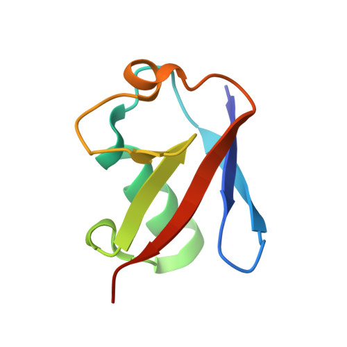
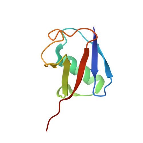
2XK5

Crystal structure of K6-linked diubiquitin


Experimental Data Snapshot

  • Method: X-RAY DIFFRACTION
  • Resolution: 3.00 Å
  • R-Value Free: 0.248 
  • R-Value Work: 0.213 
  • R-Value Observed: 0.218 

Starting Model: experimental
View more details

wwPDB Validation   3D Report Full Report


This is version 1.3 of the entry. See complete history


Literature

Engineered Diubiquitin Synthesis Reveals K29-Isopeptide Specificity of an Otu Deubiquitinase

Virdee, S.Ye, Y.Nguyen, D.P.Komander, D.Chin, J.W.

(2010) Nat Chem Biol 6: 750

  • DOI: https://doi.org/10.1038/nchembio.426
  • Primary Citation of Related Structures:  
    2XK5

  • PubMed Abstract: 

    Ubiquitination is a reversible post-translational modification that regulates a myriad of eukaryotic functions. Our ability to study the effects of ubiquitination is often limited by the inaccessibility of homogeneously ubiquitinated proteins. In particular, elucidating the roles of the so-called 'atypical' ubiquitin chains (chains other than Lys48- or Lys63-linked ubiquitin), which account for a large fraction of ubiquitin polymers, is challenging because the enzymes for their biosynthesis are unknown. Here we combine genetic code expansion, intein chemistry and chemoselective ligations to synthesize 'atypical' ubiquitin chains. We solve the crystal structure of Lys6-linked diubiquitin, which is distinct from that of structurally characterized ubiquitin chains, providing a molecular basis for the different biological functions this linkage may regulate. Moreover, we profile a panel containing 10% of the known human deubiquitinases on Lys6- and Lys29-linked ubiquitin and discover that TRABID cleaves the Lys29 linkage 40-fold more efficiently than the Lys63 linkage.


  • Organizational Affiliation

    Medical Research Council Laboratory of Molecular Biology, Cambridge, England, United Kingdom.


Macromolecules
Find similar proteins by:  (by identity cutoff)  |  3D Structure
Entity ID: 1
MoleculeChains Sequence LengthOrganismDetailsImage
UBIQUITIN76Homo sapiensMutation(s): 0 
UniProt & NIH Common Fund Data Resources
Find proteins for P62979 (Homo sapiens)
Explore P62979 
Go to UniProtKB:  P62979
PHAROS:  P62979
GTEx:  ENSG00000143947 
Entity Groups  
Sequence Clusters30% Identity50% Identity70% Identity90% Identity95% Identity100% Identity
UniProt GroupP62979
Sequence Annotations
Expand
  • Reference Sequence
Find similar proteins by:  (by identity cutoff)  |  3D Structure
Entity ID: 2
MoleculeChains Sequence LengthOrganismDetailsImage
UBIQUITIN76Homo sapiensMutation(s): 0 
UniProt & NIH Common Fund Data Resources
Find proteins for P62979 (Homo sapiens)
Explore P62979 
Go to UniProtKB:  P62979
PHAROS:  P62979
GTEx:  ENSG00000143947 
Entity Groups  
Sequence Clusters30% Identity50% Identity70% Identity90% Identity95% Identity100% Identity
UniProt GroupP62979
Sequence Annotations
Expand
  • Reference Sequence
Experimental Data & Validation

Experimental Data

  • Method: X-RAY DIFFRACTION
  • Resolution: 3.00 Å
  • R-Value Free: 0.248 
  • R-Value Work: 0.213 
  • R-Value Observed: 0.218 
  • Space Group: P 43 3 2
Unit Cell:
Length ( Å )Angle ( ˚ )
a = 104.84α = 90
b = 104.84β = 90
c = 104.84γ = 90
Software Package:
Software NamePurpose
PHENIXrefinement
iMOSFLMdata reduction
SCALAdata scaling
PHASERphasing

Structure Validation

View Full Validation Report


Entry History 

Deposition Data

Revision History  (Full details and data files)

  • Version 1.0: 2010-08-25
    Type: Initial release
  • Version 1.1: 2011-05-08
    Changes: Version format compliance
  • Version 1.2: 2011-07-13
    Changes: Version format compliance
  • Version 1.3: 2023-12-20
    Changes: Data collection, Database references, Derived calculations, Other, Refinement description