2XHG | pdb_00002xhg

Crystal Structure of the Epimerization Domain from the Initiation Module of Tyrocidine Biosynthesis


Experimental Data Snapshot

  • Method: X-RAY DIFFRACTION
  • Resolution: 1.50 Å
  • R-Value Free: 
    0.179 (Depositor), 0.197 (DCC) 
  • R-Value Work: 
    0.160 (Depositor), 0.177 (DCC) 
  • R-Value Observed: 
    0.160 (Depositor) 

wwPDB Validation 3D Report Full Report

Validation slider image for 2XHG

This is version 1.3 of the entry. See complete history

Literature

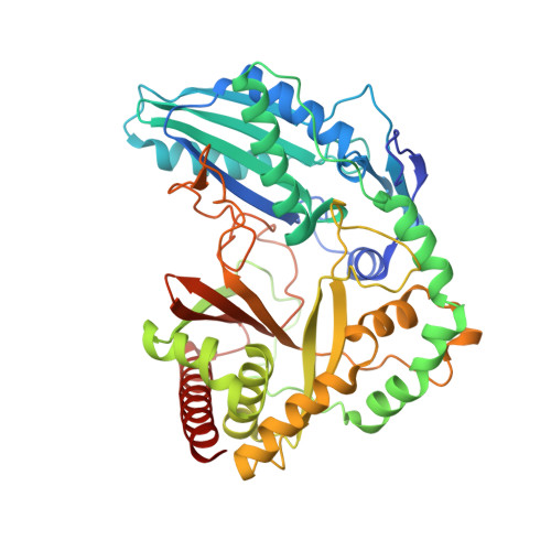
Structure of the Epimerization Domain of Tyrocidine Synthetase A

Samel, S.-A.Czodrowski, P.Essen, L.-O.

(2014) Acta Crystallogr D Biol Crystallogr 70: 1442

  • DOI: https://doi.org/10.1107/S1399004714004398
  • Primary Citation Related Structures: 
    2XHG

  • PubMed Abstract: 

    Tyrocidine, a macrocyclic decapeptide from Bacillus brevis, is nonribosomally assembled by a set of multimodular peptide synthetases, which condense two D-amino acids and eight L-amino acids to produce this membrane-disturbing antibiotic. D-Phenylalanine, the first amino acid incorporated into tyrocidine, is catalytically derived from enzyme-bound L-Phe by the C-terminal epimerization (E) domain of tyrocidine synthetase A (TycA). The 1.5 Å resolution structure of the cofactor-independent TycA E domain reveals an intimate relationship to the condensation (C) domains of peptide synthetases. In contrast to the latter, the TycA E domain uses an enlarged bridge region to plug the active-site canyon from the acceptor side, whereas at the donor side a latch-like floor loop is suitably extended to accommodate the αIII helix of the preceding peptide-carrier domain. Additionally, E domains exclusively harbour a conserved glutamate residue, Glu882, that is opposite the active-site residue His743. This active-site topology implies Glu882 as a candidate acid-base catalyst, whereas His743 stabilizes in the protonated state a transient enolate intermediate of the L↔D isomerization.


  • Organizational Affiliation
    • Biomedical Research Centre/FB15, Philipps Universität, Hans-Meerwein-Strasse 4, 35032 Marburg, Germany.

Macromolecule Content 

  • Total Structure Weight: 53.46 kDa 
  • Atom Count: 4,175 
  • Modeled Residue Count: 457 
  • Deposited Residue Count: 466 
  • Unique protein chains: 1

Macromolecules

Find similar proteins by:|  3D Structure
Entity ID: 1
MoleculeChains  Sequence LengthOrganismDetailsImage
TYROCIDINE SYNTHETASE A466Brevibacillus brevisMutation(s): 0 
UniProt
Find proteins for G1K3P2 (Brevibacillus brevis)
Explore G1K3P2 
Go to UniProtKB:  G1K3P2
Entity Groups
Sequence Clusters30% Identity50% Identity70% Identity90% Identity95% Identity100% Identity
UniProt GroupG1K3P2
Sequence Annotations
Expand
Reference Sequence

Experimental Data & Validation

Experimental Data

  • Method: X-RAY DIFFRACTION
  • Resolution: 1.50 Å
  • R-Value Free:  0.179 (Depositor), 0.197 (DCC) 
  • R-Value Work:  0.160 (Depositor), 0.177 (DCC) 
  • R-Value Observed: 0.160 (Depositor) 
Space Group: P 21 21 21
Unit Cell:
Length ( Å )Angle ( ˚ )
a = 47.71α = 90
b = 74.59β = 90
c = 124.75γ = 90
Software Package:
Software NamePurpose
REFMACrefinement
SHELXphasing

Structure Validation

View Full Validation Report


Entry History 

Deposition Data

Revision History  (Full details and data files)

  • Version 1.0: 2011-07-06
    Type: Initial release
  • Version 1.1: 2014-05-14
    Changes: Database references, Non-polymer description, Source and taxonomy, Version format compliance
  • Version 1.2: 2014-10-22
    Changes: Database references
  • Version 1.3: 2024-05-08
    Changes: Data collection, Database references, Derived calculations, Other