2XH2 | pdb_00002xh2

Engineering the enolase active site pocket: Crystal structure of the S39N D321A mutant of yeast enolase 1


Experimental Data Snapshot

  • Method: X-RAY DIFFRACTION
  • Resolution: 1.80 Å
  • R-Value Free: 
    0.209 (Depositor), 0.208 (DCC) 
  • R-Value Work: 
    0.166 (Depositor), 0.166 (DCC) 
  • R-Value Observed: 
    0.168 (Depositor) 

Starting Model: experimental
View more details

wwPDB Validation 3D Report Full Report

Validation slider image for 2XH2

Ligand Structure Quality Assessment 


This is version 1.3 of the entry. See complete history

Literature

Engineering the Enolase Magnesium II Binding Site -Implications for its Evolution.

Schreier, B.Hoecker, B.

(2010) Biochemistry 49: 7582

  • DOI: https://doi.org/10.1021/bi100954f
  • Primary Citation Related Structures: 
    2XGZ, 2XH0, 2XH2, 2XH4, 2XH7

  • PubMed Abstract: 

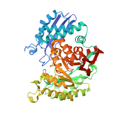
    The glycolytic enzyme enolase catalyzes the reversible elimination of water from 2-phosphoglycerate (2-PGA) to form phosphoenolpyruvate (PEP). Two magnesium ions in the active site are thought to facilitate the reaction by activation of the C2 proton of 2-PGA and charge stabilization of the intermediate. The initial abstraction of a proton from a carboxylic acid is common to all members of the enolase superfamily, yet in all other known members of this superfamily, only one magnesium ion (MgI) per active site is sufficient to promote catalysis. We wanted to further investigate the importance of the second magnesium ion (MgII) for the catalytic mechanism of yeast enolase 1. Toward this end, we removed all MgII coordinating residues and replaced substrate-MgII interactions by introducing positively charged side chains. High-resolution crystal structures and activity assays show that the introduced positively charged side chains effectively prohibit MgII binding but fail to promote catalysis. We conclude that enolase is inactive without MgII, yet control mutants without additional positively charged side chains retain basal enolase activity through binding of magnesium to 2-PGA in an open active site without the help of MgII coordinating residues. Thus, we believe that ancestral enolase activity might have evolved in a member of the enolase superfamily that provides only the necessary catalytic residues and the binding site for MgI. Additionally, precatalytic binding of 2-PGA to the apo state of enolase was observed.


  • Organizational Affiliation
    • Max Planck Institute for Developmental Biology, Spemannstrasse 35, 72076 Tübingen, Germany.

Macromolecule Content 

  • Total Structure Weight: 191.53 kDa 
  • Atom Count: 15,921 
  • Modeled Residue Count: 1,752 
  • Deposited Residue Count: 1,772 
  • Unique protein chains: 1

Macromolecules

Find similar proteins by:|  3D Structure
Entity ID: 1
MoleculeChains  Sequence LengthOrganismDetailsImage
ENOLASE 1
A, B, C, D
443Saccharomyces cerevisiaeMutation(s): 2 
EC: 4.2.1.11
UniProt
Find proteins for P00924 (Saccharomyces cerevisiae (strain ATCC 204508 / S288c))
Explore P00924 
Go to UniProtKB:  P00924
Entity Groups
Sequence Clusters30% Identity50% Identity70% Identity90% Identity95% Identity100% Identity
UniProt GroupP00924
Sequence Annotations
Expand
Reference Sequence

Small Molecules

Ligands 2 Unique
IDChains Name / Formula / InChI Key2D Diagram3D Interactions
2PG

Query on 2PG


Download:Ideal Coordinates CCD File
F [auth A],
H [auth B],
J [auth C],
L [auth D]
2-PHOSPHOGLYCERIC ACID
C3 H7 O7 P
GXIURPTVHJPJLF-UWTATZPHSA-N
MG

Query on MG


Download:Ideal Coordinates CCD File
E [auth A],
G [auth B],
I [auth C],
K [auth D]
MAGNESIUM ION
Mg
JLVVSXFLKOJNIY-UHFFFAOYSA-N

Experimental Data & Validation

Experimental Data

  • Method: X-RAY DIFFRACTION
  • Resolution: 1.80 Å
  • R-Value Free:  0.209 (Depositor), 0.208 (DCC) 
  • R-Value Work:  0.166 (Depositor), 0.166 (DCC) 
  • R-Value Observed: 0.168 (Depositor) 
Space Group: P 1
Unit Cell:
Length ( Å )Angle ( ˚ )
a = 64.72α = 89.41
b = 82.54β = 71.31
c = 95.61γ = 84.8
Software Package:
Software NamePurpose
REFMACrefinement
XDSdata reduction
XDSdata scaling
MOLREPphasing

Structure Validation

View Full Validation Report


Ligand Structure Quality Assessment 


Entry History 

Deposition Data

Revision History  (Full details and data files)

  • Version 1.0: 2010-08-25
    Type: Initial release
  • Version 1.1: 2011-05-08
    Changes: Version format compliance
  • Version 1.2: 2011-07-13
    Changes: Version format compliance
  • Version 1.3: 2023-12-20
    Changes: Data collection, Database references, Derived calculations, Other, Refinement description