2X8K | pdb_00002x8k

Crystal Structure of SPP1 Dit (gp 19.1) Protein, a Paradigm of Hub Adsorption Apparatus in Gram-positive Infecting Phages.


Experimental Data Snapshot

  • Method: X-RAY DIFFRACTION
  • Resolution: 2.95 Å
  • R-Value Free: 
    0.236 (Depositor), 0.258 (DCC) 
  • R-Value Work: 
    0.200 (Depositor), 0.222 (DCC) 
  • R-Value Observed: 
    0.202 (Depositor) 

wwPDB Validation 3D Report Full Report

Validation slider image for 2X8K

This is version 1.3 of the entry. See complete history

Literature

Crystal Structure of Bacteriophage Spp1 Distal Tail Protein (Gp 19.1): A Baseplate Hub Paradigm in Gram+ Infecting Phages.

Veesler, D.Robin, G.Lichiere, J.Auzat, I.Tavares, P.Bron, P.Campanacci, V.Cambillau, C.

(2010) J Biological Chem 285: 36666

  • DOI: https://doi.org/10.1074/jbc.M110.157529
  • Primary Citation Related Structures: 
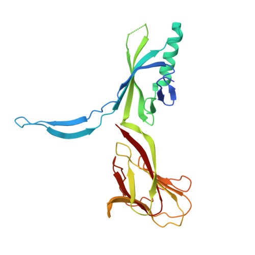
    2X8K

  • PubMed Abstract: 

    Siphophage SPP1 infects the gram-positive bacterium Bacillus subtilis using its long non-contractile tail and tail-tip. Electron microscopy (EM) previously allowed a low resolution assignment of most orf products belonging to these regions. We report here the structure of the SPP1 distal tail protein (Dit, gp19.1). The combination of x-ray crystallography, EM, and light scattering established that Dit is a back-to-back dimer of hexamers. However, Dit fitting in the virion EM maps was only possible with a hexamer located between the tail-tube and the tail-tip. Structure comparison revealed high similarity between Dit and a central component of lactophage baseplates. Sequence similarity search expanded its relatedness to several phage proteins, suggesting that Dit is a docking platform for the tail adsorption apparatus in Siphoviridae infecting gram-positive bacteria and that its architecture is a paradigm for these hub proteins. Dit structural similarity extends also to non-contractile and contractile phage tail proteins (gpV(N) and XkdM) as well as to components of the bacterial type 6 secretion system, supporting an evolutionary connection between all these devices.


  • Organizational Affiliation
    • Architecture et Fonction des Macromolécules Biologiques, UMR 6098 CNRS and Universités d'Aix-Marseille I & II, Campus de Luminy, Case 932, 13288 Marseille Cedex 09, France.

Macromolecule Content 

  • Total Structure Weight: 85.29 kDa 
  • Atom Count: 5,710 
  • Modeled Residue Count: 727 
  • Deposited Residue Count: 756 
  • Unique protein chains: 1

Macromolecules

Find similar proteins by:|  3D Structure
Entity ID: 1
MoleculeChains  Sequence LengthOrganismDetailsImage
HYPOTHETICAL PROTEIN 19.1
A, B, C
252Bacillus phage SPP1Mutation(s): 0 
UniProt
Find proteins for O48459 (Bacillus phage SPP1)
Explore O48459 
Go to UniProtKB:  O48459
Entity Groups
Sequence Clusters30% Identity50% Identity70% Identity90% Identity95% Identity100% Identity
UniProt GroupO48459
Sequence Annotations
Expand
Reference Sequence

Experimental Data & Validation

Experimental Data

  • Method: X-RAY DIFFRACTION
  • Resolution: 2.95 Å
  • R-Value Free:  0.236 (Depositor), 0.258 (DCC) 
  • R-Value Work:  0.200 (Depositor), 0.222 (DCC) 
  • R-Value Observed: 0.202 (Depositor) 
Space Group: I 2 2 2
Unit Cell:
Length ( Å )Angle ( ˚ )
a = 107.188α = 90
b = 125.745β = 90
c = 189.353γ = 90
Software Package:
Software NamePurpose
BUSTERrefinement
XDSdata reduction
SCALAdata scaling
AMoREphasing

Structure Validation

View Full Validation Report


Entry History 

Deposition Data

Revision History  (Full details and data files)

  • Version 1.0: 2010-09-15
    Type: Initial release
  • Version 1.1: 2011-05-08
    Changes: Version format compliance
  • Version 1.2: 2011-07-13
    Changes: Version format compliance
  • Version 1.3: 2024-05-08
    Changes: Data collection, Database references, Other