2X1E | pdb_00002x1e

The crystal structure of mature acyl coenzyme A:isopenicillin N acyltransferase from Penicillium chrysogenum in complex 6- aminopenicillanic acid


Experimental Data Snapshot

  • Method: X-RAY DIFFRACTION
  • Resolution: 2.00 Å
  • R-Value Free: 
    0.202 (Depositor), 0.209 (DCC) 
  • R-Value Work: 
    0.177 (Depositor), 0.188 (DCC) 
  • R-Value Observed: 
    0.178 (Depositor) 

Starting Model: experimental
View more details

wwPDB Validation 3D Report Full Report

Validation slider image for 2X1E

Ligand Structure Quality Assessment 


This is version 1.5 of the entry. See complete history

Literature

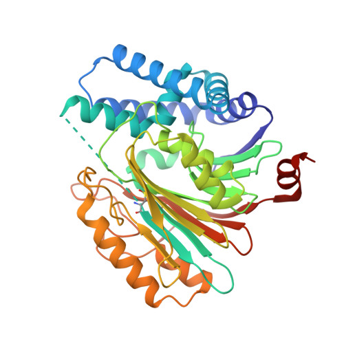
Structures of an Isopenicillin N Converting Ntn-Hydrolase Reveal Different Catalytic Roles for the Active Site Residues of Precursor and Mature Enzyme.

Bokhove, M.Yoshida, H.Hensgens, C.M.H.Van Der Laan, J.M.Sutherland, J.D.Dijkstra, B.W.

(2010) Structure 18: 301

  • DOI: https://doi.org/10.1016/j.str.2010.01.005
  • Primary Citation Related Structures: 
    2X1C, 2X1D, 2X1E

  • PubMed Abstract: 

    Penicillium chrysogenum Acyl coenzyme A:isopenicillin N acyltransferase (AT) performs the last step in the biosynthesis of hydrophobic penicillins, exchanging the hydrophilic side chain of a precursor for various hydrophobic side chains. Like other N-terminal nucleophile hydrolases AT is produced as an inactive precursor that matures upon posttranslational cleavage. The structure of a Cys103Ala precursor mutant shows that maturation is autoproteolytic, initiated by Cys103 cleaving its preceding peptide bond. The crystal structure of the mature enzyme shows that after autoproteolysis residues 92-102 fold outwards, exposing a buried pocket. This pocket is structurally and chemically flexible and can accommodate substrates of different size and polarity. Modeling of a substrate-bound state indicates the residues important for catalysis. Comparison of the proposed autoproteolytic and substrate hydrolysis mechanisms shows that in both events the same catalytic residues are used, but that they perform different roles in catalysis.


  • Organizational Affiliation
    • Laboratory of Biophysical Chemistry, University of Groningen, Nijenborgh 4, Groningen, Netherlands.

Macromolecule Content 

  • Total Structure Weight: 161.27 kDa 
  • Atom Count: 11,691 
  • Modeled Residue Count: 1,407 
  • Deposited Residue Count: 1,428 
  • Unique protein chains: 1

Macromolecules

Find similar proteins by:|  3D Structure
Entity ID: 1
MoleculeChains  Sequence LengthOrganismDetailsImage
ACYL-COENZYME
A, B, C, D
357Penicillium chrysogenumMutation(s): 0 
EC: 2.3.1.164
UniProt
Find proteins for P15802 (Penicillium chrysogenum)
Explore P15802 
Go to UniProtKB:  P15802
Entity Groups
Sequence Clusters30% Identity50% Identity70% Identity90% Identity95% Identity100% Identity
UniProt GroupP15802
Sequence Annotations
Expand
Reference Sequence

Small Molecules

Ligands 4 Unique
IDChains Name / Formula / InChI Key2D Diagram3D Interactions
X1E

Query on X1E


Download:Ideal Coordinates CCD File
G [auth A](2S,5R,6R)-6-AMINO-3,3-DIMETHYL-7-OXO-4-THIA-1-AZABICYCLO[3.2.0]HEPTANE-2-CARBOXYLIC ACID
C8 H12 N2 O3 S
NGHVIOIJCVXTGV-ALEPSDHESA-N
PO4

Query on PO4


Download:Ideal Coordinates CCD File
E [auth A],
I [auth B],
O [auth D]
PHOSPHATE ION
O4 P
NBIIXXVUZAFLBC-UHFFFAOYSA-K
GOL

Query on GOL


Download:Ideal Coordinates CCD File
F [auth A]
H [auth A]
K [auth C]
L [auth C]
M [auth C]
F [auth A],
H [auth A],
K [auth C],
L [auth C],
M [auth C],
N [auth C],
P [auth D]
GLYCEROL
C3 H8 O3
PEDCQBHIVMGVHV-UHFFFAOYSA-N
CL

Query on CL


Download:Ideal Coordinates CCD File
J [auth B]CHLORIDE ION
Cl
VEXZGXHMUGYJMC-UHFFFAOYSA-M
Modified Residues  1 Unique
IDChains TypeFormula2D DiagramParent
CSD
Query on CSD
A, B, C, D
L-PEPTIDE LINKINGC3 H7 N O4 SCYS

Experimental Data & Validation

Experimental Data

  • Method: X-RAY DIFFRACTION
  • Resolution: 2.00 Å
  • R-Value Free:  0.202 (Depositor), 0.209 (DCC) 
  • R-Value Work:  0.177 (Depositor), 0.188 (DCC) 
  • R-Value Observed: 0.178 (Depositor) 
Space Group: C 1 2 1
Unit Cell:
Length ( Å )Angle ( ˚ )
a = 198.18α = 90
b = 68.38β = 128.81
c = 146.57γ = 90
Software Package:
Software NamePurpose
REFMACrefinement
XDSdata reduction
SCALAdata scaling
MOLREPphasing

Structure Validation

View Full Validation Report


Ligand Structure Quality Assessment 


Entry History 

Deposition Data

Revision History  (Full details and data files)

  • Version 1.0: 2010-03-09
    Type: Initial release
  • Version 1.1: 2011-12-28
    Changes: Database references, Derived calculations, Non-polymer description, Refinement description, Version format compliance
  • Version 1.2: 2014-11-05
    Changes: Atomic model, Derived calculations, Non-polymer description, Other, Structure summary
  • Version 1.3: 2019-07-24
    Changes: Data collection, Derived calculations
  • Version 1.4: 2023-12-20
    Changes: Data collection, Database references, Derived calculations, Other, Refinement description
  • Version 1.5: 2024-11-13
    Changes: Structure summary