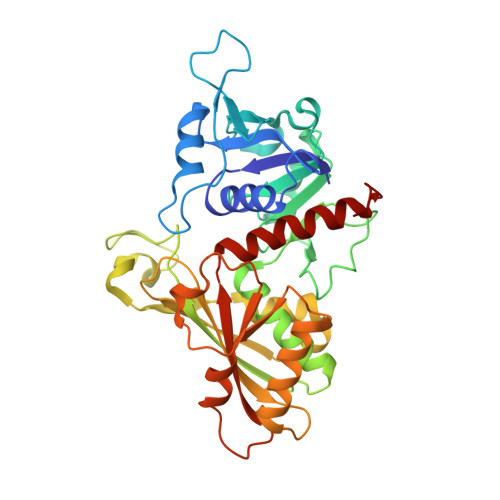
2X0N | pdb_00002x0n

Structure of glycosomal glyceraldehyde-3-phosphate dehydrogenase from Trypanosoma brucei determined from Laue data


Experimental Data Snapshot

  • Method: X-RAY DIFFRACTION
  • Resolution: 3.20 Å
  • R-Value Free: 
    0.218 (Depositor), 0.220 (DCC) 
  • R-Value Work: 
    0.150 (Depositor), 0.150 (DCC) 
  • R-Value Observed: 
    0.153 (Depositor) 

Starting Model: experimental
View more details

wwPDB Validation   3D Report Full Report


Ligand Structure Quality Assessment 

Created with Raphaël 2.3.0Worse 01 BetterLigand structure goodness of fit to experimental dataBest fitted NADClick on this verticalbar to view details

This is version 1.2 of the entry. See complete history


Literature

Structure of Glycosomal Glyceraldehyde-3-Phosphate Dehydrogenase from Trypanosoma Brucei Determined from Laue Data.

Vellieux, F.M.D.Hajdu, J.Verlinde, C.L.Groendijk, H.Read, R.J.Greenhough, T.J.Campbell, J.W.Kalk, K.H.Littlechild, J.A.Watson, H.C.

(1993) Proc Natl Acad Sci U S A 90: 2355

  • DOI: https://doi.org/10.1073/pnas.90.6.2355
  • Primary Citation of Related Structures:  
    2X0N

  • PubMed Abstract: 

    The three-dimensional structure of glycosomal glyceraldehyde-3-phosphate dehydrogenase [D-glyceraldehyde-3-phosphate:NAD+ oxidoreductase (phosphorylating), EC 1.12.1.12] from the sleeping-sickness parasite Trypanosoma brucei was solved by molecular replacement at 3.2-A resolution with an x-ray data set collected by the Laue method. For data collection, three crystals were exposed to the polychromatic synchrotron x-ray beam for a total of 20.5 sec. The structure was solved by using the Bacillus stearothermophilus enzyme model [Skarzyński, T., Moody, P. C. E. & Wonacott, A. J. (1987) J. Mol. Biol. 193, 171-187] with a partial data set which was 37% complete. The crystals contain six subunits per asymmetric unit, which allowed us to overcome the absence of > 60% of the reflections by 6-fold density averaging. After molecular dynamics refinement, the current molecular model has an R factor of 17.6%. Comparing the structure of the trypanosome enzyme with that of the homologous human muscle enzyme, which was determined at 2.4-A resolution, reveals important structural differences in the NAD binding region. These are of great interest for the design of specific inhibitors of the parasite enzyme.


  • Organizational Affiliation

    Department of Chemistry, BIOSON Research Institute, University of Groningen, The Netherlands.


Macromolecules
Find similar proteins by:  (by identity cutoff)  |  3D Structure
Entity ID: 1
MoleculeChains Sequence LengthOrganismDetailsImage
GLYCERALDEHYDE-3-PHOSPHATE DEHYDROGENASE, GLYCOSOMAL359Trypanosoma brucei bruceiMutation(s): 0 
EC: 1.2.1.12
UniProt
Find proteins for P22512 (Trypanosoma brucei brucei)
Explore P22512 
Go to UniProtKB:  P22512
Entity Groups  
Sequence Clusters30% Identity50% Identity70% Identity90% Identity95% Identity100% Identity
UniProt GroupP22512
Sequence Annotations
Expand
  • Reference Sequence
Small Molecules
Ligands 2 Unique
IDChains Name / Formula / InChI Key2D Diagram3D Interactions
NAD
Query on NAD

Download Ideal Coordinates CCD File 
I [auth A]
L [auth B]
O
R [auth P]
U [auth Q]
NICOTINAMIDE-ADENINE-DINUCLEOTIDE
C21 H27 N7 O14 P2
BAWFJGJZGIEFAR-NNYOXOHSSA-N
SO4
Query on SO4

Download Ideal Coordinates CCD File 
G [auth A]
H [auth A]
J [auth B]
K [auth B]
M [auth O]
SULFATE ION
O4 S
QAOWNCQODCNURD-UHFFFAOYSA-L
Experimental Data & Validation

Experimental Data

  • Method: X-RAY DIFFRACTION
  • Resolution: 3.20 Å
  • R-Value Free:  0.218 (Depositor), 0.220 (DCC) 
  • R-Value Work:  0.150 (Depositor), 0.150 (DCC) 
  • R-Value Observed: 0.153 (Depositor) 
Space Group: P 21 21 2
Unit Cell:
Length ( Å )Angle ( ˚ )
a = 135.52α = 90
b = 256.27β = 90
c = 114.91γ = 90
Software Package:
Software NamePurpose
PHENIXrefinement
DARESBURYdata reduction
DARESBURYdata scaling
MERLOTphasing
BIOMOLphasing

Structure Validation

View Full Validation Report


Ligand Structure Quality Assessment 

Created with Raphaël 2.3.0Worse 01 BetterLigand structure goodness of fit to experimental dataBest fitted NADClick on this verticalbar to view details

Entry History 

Deposition Data

Revision History  (Full details and data files)

  • Version 1.0: 2009-12-22
    Type: Initial release
  • Version 1.1: 2017-01-25
    Changes: Data collection
  • Version 1.2: 2023-12-20
    Changes: Data collection, Database references, Derived calculations, Other, Refinement description