2WYO | pdb_00002wyo

Trypanosoma brucei glutathione synthetase


Experimental Data Snapshot

  • Method: X-RAY DIFFRACTION
  • Resolution: 3.15 Å
  • R-Value Free: 
    0.277 (Depositor), 0.276 (DCC) 
  • R-Value Work: 
    0.203 (Depositor), 0.202 (DCC) 
  • R-Value Observed: 
    0.206 (Depositor) 

Starting Model: experimental
View more details

wwPDB Validation 3D Report Full Report

Validation slider image for 2WYO

Ligand Structure Quality Assessment 


This is version 1.3 of the entry. See complete history

Literature

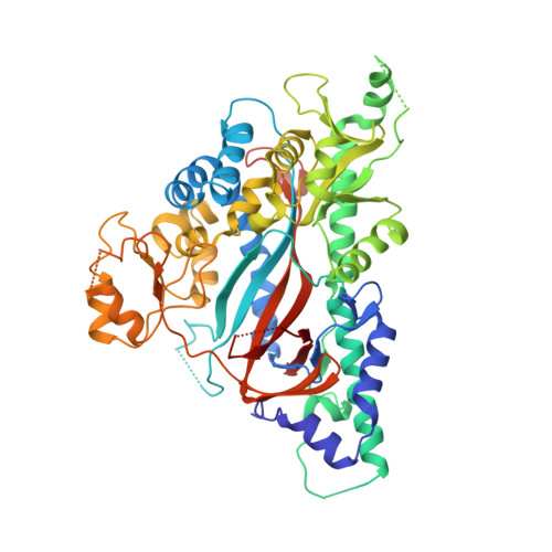
Structure of Trypanosoma Brucei Glutathione Synthetase; Domain and Loop Alterations in the Catalytic Cycle of a Highly Conserved Enzyme.

Fyfe, P.K.Alphey, M.S.Hunter, W.N.

(2010) Mol Biochem Parasitol 170: 93

  • DOI: https://doi.org/10.1016/j.molbiopara.2009.12.011
  • Primary Citation Related Structures: 
    2WYO

  • PubMed Abstract: 

    Glutathione synthetase catalyses the synthesis of the low molecular mass thiol glutathione from l-gamma-glutamyl-l-cysteine and glycine. We report the crystal structure of the dimeric enzyme from Trypanosoma brucei in complex with the product glutathione. The enzyme belongs to the ATP-grasp family, a group of enzymes known to undergo conformational changes upon ligand binding. The T. brucei enzyme crystal structure presents two dimers in the asymmetric unit. The structure reveals variability in the order and position of a small domain, which forms a lid for the active site and serves to capture conformations likely to exist during the catalytic cycle. Comparisons with orthologous enzymes, in particular from Homo sapiens and Saccharomyces cerevisae, indicate a high degree of sequence and structure conservation in part of the active site. Structural differences that are observed between the orthologous enzymes are assigned to different ligand binding states since key residues are conserved. This suggests that the molecular determinants of ligand recognition and reactivity are highly conserved across species. We conclude that it would be difficult to target the parasite enzyme in preference to the host enzyme and therefore glutathione synthetase may not be a suitable target for antiparasitic drug discovery.


  • Organizational Affiliation
    • Division of Biological Chemistry and Drug Discovery, College of Life Sciences, University of Dundee, Dow Street, Dundee, DD1 5EH, United Kingdom.

Macromolecule Content 

  • Total Structure Weight: 250.83 kDa 
  • Atom Count: 15,702 
  • Modeled Residue Count: 1,996 
  • Deposited Residue Count: 2,248 
  • Unique protein chains: 1

Macromolecules

Find similar proteins by:|  3D Structure
Entity ID: 1
MoleculeChains  Sequence LengthOrganismDetailsImage
GLUTATHIONE SYNTHETASE
A, B, C, D
562Trypanosoma bruceiMutation(s): 0 
EC: 6.3.2.3
UniProt
Find proteins for Q57UN0 (Trypanosoma brucei brucei (strain 927/4 GUTat10.1))
Explore Q57UN0 
Go to UniProtKB:  Q57UN0
Entity Groups
Sequence Clusters30% Identity50% Identity70% Identity90% Identity95% Identity100% Identity
UniProt GroupQ57UN0
Sequence Annotations
Expand
Reference Sequence

Small Molecules

Ligands 2 Unique
IDChains Name / Formula / InChI Key2D Diagram3D Interactions
GSH

Query on GSH


Download:Ideal Coordinates CCD File
E [auth A],
H [auth B],
J [auth C],
N [auth D]
Glutathione
C10 H17 N3 O6 S
RWSXRVCMGQZWBV-WDSKDSINSA-N
SO4

Query on SO4


Download:Ideal Coordinates CCD File
F [auth A]
G [auth A]
I [auth B]
K [auth C]
L [auth C]
F [auth A],
G [auth A],
I [auth B],
K [auth C],
L [auth C],
M [auth C],
O [auth D]
SULFATE ION
O4 S
QAOWNCQODCNURD-UHFFFAOYSA-L

Experimental Data & Validation

Experimental Data

  • Method: X-RAY DIFFRACTION
  • Resolution: 3.15 Å
  • R-Value Free:  0.277 (Depositor), 0.276 (DCC) 
  • R-Value Work:  0.203 (Depositor), 0.202 (DCC) 
  • R-Value Observed: 0.206 (Depositor) 
Space Group: P 21 21 21
Unit Cell:
Length ( Å )Angle ( ˚ )
a = 92.59α = 90
b = 125.15β = 90
c = 243.17γ = 90
Software Package:
Software NamePurpose
REFMACrefinement
XDSdata reduction
XSCALEdata scaling
PHASERphasing

Structure Validation

View Full Validation Report


Ligand Structure Quality Assessment 


Entry History 

Deposition Data

Revision History  (Full details and data files)

  • Version 1.0: 2010-01-19
    Type: Initial release
  • Version 1.1: 2011-07-13
    Changes: Advisory, Refinement description, Version format compliance
  • Version 1.2: 2011-12-07
    Changes: Atomic model, Derived calculations, Non-polymer description, Other, Refinement description, Source and taxonomy
  • Version 1.3: 2023-12-20
    Changes: Data collection, Database references, Derived calculations, Other, Refinement description