2WUL

CRYSTAL STRUCTURE OF THE HUMAN GLUTAREDOXIN 5 WITH BOUND GLUTATHIONE IN AN FES CLUSTER


Experimental Data Snapshot

  • Method: X-RAY DIFFRACTION
  • Resolution: 2.40 Å
  • R-Value Free: 0.251 
  • R-Value Work: 0.200 
  • R-Value Observed: 0.202 

Starting Model: experimental
View more details

wwPDB Validation   3D Report Full Report


Ligand Structure Quality Assessment 


This is version 1.4 of the entry. See complete history


Literature

The Crystal Structure of Human Glrx5: Iron Sulphur Cluster Coordination, Tetrameric Assembly and Monomer Activity.

Johansson, C.Roos, A.K.Montano, S.J.Sengupta, R.Filippakopoulos, P.Guo, K.von Delft, F.Holmgren, A.Oppermann, U.Kavanagh, K.L.

(2011) Biochem J 433: 303

  • DOI: https://doi.org/10.1042/BJ20101286
  • Primary Citation of Related Structures:  
    2WUL

  • PubMed Abstract: 

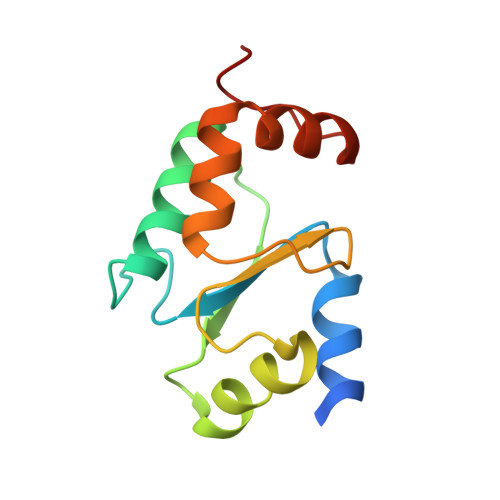
    Human GLRX5 (glutaredoxin 5) is an evolutionarily conserved thiol-disulfide oxidoreductase that has a direct role in the maintenance of normal cytosolic and mitochondrial iron homoeostasis, and its expression affects haem biosynthesis and erythropoiesis. We have crystallized the human GLRX5 bound to two [2Fe-2S] clusters and four GSH molecules. The crystal structure revealed a tetrameric organization with the [2Fe-2S] clusters buried in the interior and shielded from the solvent by the conserved β1-α2 loop, Phe⁶⁹ and the GSH molecules. Each [2Fe-2S] cluster is ligated by the N-terminal activesite cysteine (Cys⁶⁷) thiols contributed by two protomers and two cysteine thiols from two GSH. The two subunits co-ordinating the cluster are in a more extended conformation compared with iron-sulfur-bound human GLRX2, and the intersubunit interactions are more extensive and involve conserved residues among monothiol GLRXs. Gel-filtration chromatography and analytical ultracentrifugation support a tetrameric organization of holo-GLRX5, whereas the apoprotein is monomeric. MS analyses revealed glutathionylation of the cysteine residues in the absence of the [2Fe-2S] cluster, which would protect them from further oxidation and possibly facilitate cluster transfer/acceptance. Apo-GLRX5 reduced glutathione mixed disulfides with a rate 100 times lower than did GLRX2 and was active as a glutathione-dependent electron donor for mammalian ribonucleotide reductase.


  • Organizational Affiliation

    University of Oxford, UK. catrine.johansson@sgc.ox.ac.uk


Macromolecules
Find similar proteins by:  (by identity cutoff)  |  3D Structure
Entity ID: 1
MoleculeChains Sequence LengthOrganismDetailsImage
GLUTAREDOXIN RELATED PROTEIN 5
A, B, C, D
118Homo sapiensMutation(s): 0 
UniProt & NIH Common Fund Data Resources
Find proteins for Q86SX6 (Homo sapiens)
Explore Q86SX6 
Go to UniProtKB:  Q86SX6
GTEx:  ENSG00000182512 
Entity Groups  
Sequence Clusters30% Identity50% Identity70% Identity90% Identity95% Identity100% Identity
UniProt GroupQ86SX6
Sequence Annotations
Expand
  • Reference Sequence
Small Molecules
Ligands 4 Unique
IDChains Name / Formula / InChI Key2D Diagram3D Interactions
GSH
Query on GSH

Download Ideal Coordinates CCD File 
E [auth A],
G [auth B],
J [auth C],
L [auth D]
GLUTATHIONE
C10 H17 N3 O6 S
RWSXRVCMGQZWBV-WDSKDSINSA-N
FES
Query on FES

Download Ideal Coordinates CCD File 
F [auth A],
K [auth C]
FE2/S2 (INORGANIC) CLUSTER
Fe2 S2
NIXDOXVAJZFRNF-UHFFFAOYSA-N
PEG
Query on PEG

Download Ideal Coordinates CCD File 
I [auth B]DI(HYDROXYETHYL)ETHER
C4 H10 O3
MTHSVFCYNBDYFN-UHFFFAOYSA-N
CL
Query on CL

Download Ideal Coordinates CCD File 
H [auth B],
M [auth D],
N [auth D],
O [auth D]
CHLORIDE ION
Cl
VEXZGXHMUGYJMC-UHFFFAOYSA-M
Experimental Data & Validation

Experimental Data

  • Method: X-RAY DIFFRACTION
  • Resolution: 2.40 Å
  • R-Value Free: 0.251 
  • R-Value Work: 0.200 
  • R-Value Observed: 0.202 
  • Space Group: P 43 21 2
Unit Cell:
Length ( Å )Angle ( ˚ )
a = 68.995α = 90
b = 68.995β = 90
c = 229.331γ = 90
Software Package:
Software NamePurpose
MOSFLMdata reduction
SCALAdata scaling
SHELXphasing
SHARPphasing
REFMACrefinement

Structure Validation

View Full Validation Report


Ligand Structure Quality Assessment 


Entry History 

Revision History  (Full details and data files)

  • Version 1.0: 2009-10-20
    Type: Initial release
  • Version 1.1: 2011-07-13
    Changes: Advisory, Version format compliance
  • Version 1.2: 2011-12-07
    Changes: Atomic model, Database references, Derived calculations, Non-polymer description
  • Version 1.3: 2018-01-24
    Changes: Database references, Structure summary
  • Version 1.4: 2023-12-20
    Changes: Data collection, Database references, Derived calculations, Other, Refinement description